|
Münster (upm).
A “molecular light-switch” in the cell membrane causes the cell to take on a burr-cell form (echinocyte, r.) or a flat form (similar to a discocyte), regardless of whether it is subjected to radiation with visual light or with ultraviolet light.<address>© Adapted from Höglsperger F. et al., Nature Communications, DOI: 10.1038/s41467-023-39032-0; licensed under CC BY licence.</address>
A “molecular light-switch” in the cell membrane causes the cell to take on a burr-cell form (echinocyte, r.) or a flat form (similar to a discocyte), regardless of whether it is subjected to radiation with visual light or with ultraviolet light.
© Adapted from Höglsperger F. et al., Nature Communications, DOI: 10.1038/s41467-023-39032-0; licensed under CC BY licence.

Research team shows how a cell’s form can be reversed

Light-switchable molecules in membranes enable different forms of living cells to be studied /Study published in Nature Communications

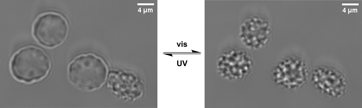
Membranes fulfil a variety of tasks in living cells: for example, they separate the cells from their surroundings and thus protect them. Also, by means of transport proteins they convey the necessary nutrients to the interior. Membranes also play a major role when cells grow together to form tissue, when they proliferate by dividing, or when they move. A team of researchers led by Prof. Bart Jan Ravoo from the Institute of Organic Chemistry at the University of Münster, and by Prof. Timo Betz from the Third Institute of Physics – Biophysics at the University of Göttingen, are now the first to describe how living cells can have their shape reversed through targeting the cell membrane by means of light. The study has been published in the journal Nature Communications.

Microscopic image of the red corpuscles with azobenzene in the membrane. The echinocyte form (l.) is observed when the photo-switch is trans-oriented, and the form similar to discocytes is observed when the photo-switch is cis-oriented.<address>© Adapted from Höglsperger F. et al., Nature Communications, DOI: 10.1038/s41467-023-39032-0; licensed under CC BY licence.</address>
Microscopic image of the red corpuscles with azobenzene in the membrane. The echinocyte form (l.) is observed when the photo-switch is trans-oriented, and the form similar to discocytes is observed when the photo-switch is cis-oriented.
© Adapted from Höglsperger F. et al., Nature Communications, DOI: 10.1038/s41467-023-39032-0; licensed under CC BY licence.
In a laboratory experiment, the team succeeded in changing the shape of red blood corpuscles. These cells are normally disc-shaped (“discocytes”), but they can also take on a prickly form with projections on the surface (“echinocytes” or “burr cells”). For their experiment, the researchers used a kind of “molecular light-switch” which they inserted into the cell membranes and which had the effect there of making the cells take on the thorny burr-cell shape. When subjected to ultraviolet light radiation, the cells changed their shape again within seconds and then looked like the flat discocytes which occur in nature. Under visual light, they again assumed their burr-cell shape. This process was repeated several times.

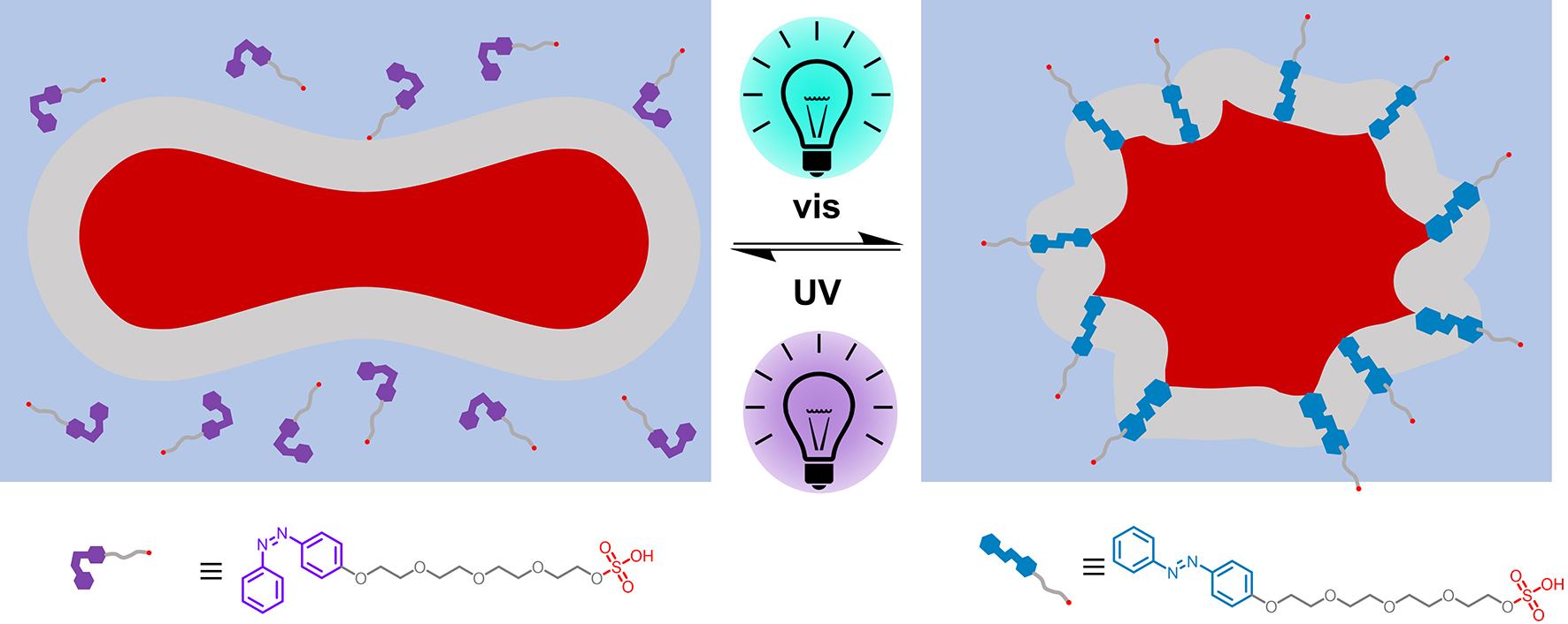
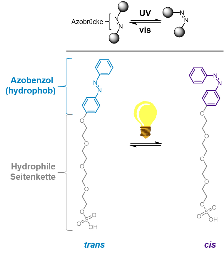
The basis for this change of shape is a molecule which has a similar structure to the molecules in the cell membrane and which has an additional functional unit which responds to light by changing its form: an azobenzene. “Because the azobenzene is hydrophobic – in other words, has water-repellent properties – we additionally inserted a hydrophilic, water-soluble side chain,” explains lead author Dr. Fabian Höglsperger from Bart Jan Ravoo’s team. “This is a design which comes very close to the lipid molecules found in nature in cell membranes. The differences between the azobenzene derivative, which we designed and produced, and the lipid molecules found in nature are small – but they are enough to cause a significant change in the cell membrane by means of light.”

An amphiphilic azobenzene is the central molecule in the study. It can be switched back and forth between two states by means of light, thus changing its properties. Also, it combines hydrophilic and hydrophobic properties in the same molecule, which makes it compatible with membranes.<address>© Münster University - Henning Klaasen</address>
An amphiphilic azobenzene is the central molecule in the study. It can be switched back and forth between two states by means of light, thus changing its properties. Also, it combines hydrophilic and hydrophobic properties in the same molecule, which makes it compatible with membranes.
© Münster University - Henning Klaasen
The team’s explanation for the fact that the cells promptly change into the flat form when subjected to ultraviolet light is the changed structure of the azobenzene molecule. There is less likelihood of this changed molecule variant residing in the cell membrane, and the projections occurring in a membrane with the echinocyte form become flatter. When subjected to visual light, the reverse reaction is triggered in the photo-switch, and the echinocyte form regresses.

The study in question deals with basic research. But, as Bart Jan Ravoo says, “In future, this simple but efficient method could help in studying the reactions of cells to their surroundings – depending on their shape – or in using light to control processes such as cell division and cell migration.”

Background details: The change of shape occurring in cells in nature is also due to the structure of the membranes. The double layer of molecules with a hydrophilic head and a hydrophobic side chain is so stable that molecules cannot readily pass through. At the same time, this layer is moveable to enable it to respond to internal and external stimuli. “As is often the case,” says Fabian Höglsperger, “this change of shape is a process which occurs easily in nature but which is difficult to control under laboratory conditions.”

The study brought together researchers from four institutes at the Universities of Münster and Göttingen – with their expertise in the fields of synthetic, theoretical and physical chemistry, as well as biophysics and cell biology. The methods used by the team in their work included spectroscopy and microscopy. The red blood corpuscles were human cells.

For their study, the researchers received financial support from the German Research Foundation as part of Collaborative Research Centres 1459 (Intelligent matter) and 1348 (Dynamic cellular interfaces), as well as from the European Research Council (ERC Grant PolarizeMe 771201).


Original publication

Fabian Höglsperger, Bart E. Vos, Arne D. Hofemeier, Maximilian D. Seyfried, Bastian Stövesand, Azadeh Alavizargar, Leon Topp, Andreas Heuer, Timo Betz, Bart Jan Ravoo (2023): Rapid and reversible optical switching of cell membrane area by an amphiphilic azobenzene. Nature Communications 14, 3760 DOI: 10.1038/s41467-023-39032-0

Further information