|
|
|
Free Neuropathology 5:30 (2024) |
|
Meeting Abstracts |
|
Northern Lights Neuroscience Symposium 2024 |
|
Expanding Spectrum of Common Dementia Disorders |
|
Meeting Abstracts |
|
|
|
|
|
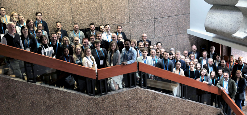
The Northern Lights Neuroscience Symposium 2024 “Expanding Spectrum of Common Dementia Disorders” was held in Hanasaari, Helsinki (Espoo), Finland on September 26–27, 2024. The meeting was jointly organised by the Scandinavian Neuropathological Society (chair Olivera Casar-Borota) and University of Helsinki. Drs. Liisa Myllykangas (chair), Olli Tynninen, Maria Gardberg and Tuomas Rauramaa made up the organising committee. The event brought together neuropathologists, clinicians and neuroscientists from nine different counties. In total, 91 people had registered for the meeting, and there were 29 abstracts. The keynote lectures on neuropathological and clinical aspects of new dementia entities such as LATE and PART as well as subtypes of AD and LBD, vascular and mixed patologies, were given by Irina Alafuzoff, Peter Nelson, David Wolk, Gabor Kovacs, Melissa Murray, Jacob Vogel, Liisa Myllykangas, Per Borghammer, Johannes Attems and Masafumi Ihara. The Patrick Sourander lecture “Investigating choroid plexus and cerebrospinal fluid roles and functions in neurologic disease“ was presented by Maria Lehtinen from Harvard Medical School. Seven additional oral presentations (selected from submitted abstracts) were also given. The participants were highly appreciative of the meeting’s top level scientific content and of the networking event, which included a Finnish sauna, a cruise in the Helsinki archipelago and a dinner in the center of Helsinki.
|
|
|
|
Keywords: Scandinavian Neuropathological Society, Meeting abstracts, Northern Lights Neuroscience Symposium 2024 |
|
Meeting Abstracts
Free Neuropathol 5:30:5 Expanding spectrum of dementia disorders Irina Alafuzoff1
During the last 40–50 years our knowledge regarding aging related cognitive impairment has evolved significantly. In 80s clinicians primarily referred to a senile dementia, a malady of the aged. The cause of this disease was mostly unknown. First International Conference on Alzheimer's Disease and Related Disorders was organized by professors Khalid Iqbal, Henry Wisniewski and Bengt Winblad in 1988 in Las Vegas, USA. The total number of participants was quite sparse, most being clinicians and neuropathologist. At this time from the view of a neuropathologist the primary topics discussed were 1. How many plaques are needed for a diagnosis of Alzheimer’s disease and 2. Which is the primary lesion seen in the brain, amyloid β-protein or hyperphosphorylated τ? Currently, several different conferences are organized yearly with thousands of participants from various fields of neuroscience. Our knowledge regarding the neuropathology seen in subjects with aging related cognitive impairment has evolved significantly. This has been facilitated by the identification of proteins that are altered in the brain of aged. The insight that these protein alterations are initially seen in certain brain regions and progress following certain neuroanatomical connections has broadened our understanding. It has also been acknowledged that in some cases the disease is inherited based on genetic alterations leading to ample use of transgenic animals in research. Sadly, we still are quite far from curative treatments of these maladies. Recently it has become clear that in many aged, not few but rather several protein alterations are to be found in the brain and even this tendency seems to increase with age. A major obstacle today is limited number of human brain tissue available for research. The autopsy frequencies are extremely low and moreover the brain is seldom assessed postmortem. Many studies can certainly be carried out in animal models. However, in line with the fruitful research carried out on tumors, where the use of surgical human samples has led to substantial results, also neurodegeneration needs to be studied in human setting. This is certainly needed to decrypt these complex diseases and hopefully find efficient treatment strategies improving the life quality of the affected subjects.
Free Neuropathol 5:30:6 RT-QuIC with universal control fluid: standardized detection of alpha-synucleinopathies Remarh Bsoul1, Eva Løbner Lund1, Kristian Steen Frederiksen1, Sara Brynhild Winther Bech2, Anja Hviid Simonsen1, Kirsten Svenstrup2, Aušrinė Areškevičiūtė1
We present a CSF RT-QuIC-αSyn protocol that is relatively easy to adapt, performs stably, and enables a uniform preparation of both sample and control reactions. We adopted and modified RT-QuIC-αSyn method described by others. Our protocol's sensitivity and specificity was estimated by testing CSF samples from Parkinson´s (PD) (N = 12), Dementia with Lewy-body (DLB) (N = 12), Alzheimer´s (AD) (N = 12), Motor Neuron Disease (MND) (N = 15), Multiple System Atrophy (MSA) (N = 18) and healthy controls (HC) (N = 12). We tested 3 volumes of each CSF sample in quadruplicates to assess the correlation between added to the reaction CSF volume and αSyn seeding efficiency. Furthermore, we developed a Universal Control Fluid (UCF) to standardize RT-QuIC reaction environment across all samples and controls by creating and testing various UCF compositions and sample/control reaction settings. Our RT-QuIC-αSyn performs with > 95 % sensitivity for PD and DLB, > 95 % specificity when compared to AD and MND, and 100 % specificity when compared to NC; whereas αSyn in MSA samples was undetectable. We have defined a UCF composition that does not compromise RT-QuIC reactions' effectivity and gives 100 % sensitivity and specificity when comparing unseeded UCF and UCF seeded with patient material. The UCF has also been tested as a CSF diluent and is suitable for broadening the seeding detection range while keeping the assay environment uniform. RT-QuIC-αSyn is a highly sensitive and specific clinical biomarker providing PD/DLB diagnosis within 48 hours, and distinguishing it from MSA. RT-QuIC-αSyn with UCF has the potential for a standardized diagnostic protocol across laboratories.
Free Neuropathol 5:30:7 Mixed pathologies of the ageing brain Johannes Attems1
The defining neuropathological features of age associated neurodegenerative diseases are aggregations of misfolded proteins and the neuropathological diagnosis is based on the semiquantitative assessment of these misfolded proteins that constitute the neuropathological hallmark lesion for the respective disease: e.g. Alzheimer's disease (AD), amyloid-β (Aβ) hyperphosphorylated tau (tau); Lewy body diseases, α-synuclein (α-syn); limbic-predominant age-related TDP-43 encephalopathy (LATE), TDP-43; frontotemporal lobar degeneration, tau or TDP-43 or ubiquitin or FUS. In addition, cerebrovascular lesions are assessed for the diagnosis of cerebrovascular disease. However, in brains of elderly patients suffering from neurodegenerative diseases mixed pathologies are usually present and various amounts of neurodegenerative and cerebrovascular pathology are frequently seen even in brains of non-demented elderly. It does indeed become increasingly clear that the clinical picture of dementia in most aged patients results from mixed pathologies rather than from one single disease. Importantly, these mixed pathologies are often not detected clinically, which has a detrimental impact on clinical studies since apparently homogeneous study cohorts (e.g. AD) are likely to be heterogeneous (e.g. AD only, AD and α-syn, AD and TDP-43), which in turn introduces a bias into therapeutic trials and biomarker/ imaging studies. Hence, wherever possible clinical studies should ideally involve neuropathological post mortem assessment to correlate clinical with neuropathological data as this will enable a more accurate stratification of clinical cohorts according to the presence of multiple pathologies. This is crucial for a meaningful interpretation of clinical data on biomarkers and therapeutic effects.
Free Neuropathol 5:30:8 Nigral neuroinflammation and dopaminergic neurons in PD, MSA and PSP: a comparative clinicopathological study Emmilotta Backman1,2, Maria Gardberg3, Laura Luntamo1,2, Markus Peurla4, Tero Vahlberg5, Per Borghammer6,7, Nadia Stefanova8, Gregor Wenning8, Valtteri Kaasinen1,2
Objective: To understand the complex interplay between neuroinflammation and clinical characteristics in Parkinson’s disease (PD), multiple system atrophy (MSA) and progressive supranuclear palsy (PSP). Materials and methods: Postmortem neuropathological samples were obtained from 79 individuals (PD, n = 38; PSP, n = 15; MSA, n = 14; and controls, n = 12). The numbers and densities of SNc tyrosine hydroxylase (TH)-positive neurons and T cells (CD3+, CD4+ and CD8+) and Iba1 expression (a microglial marker) were assessed in both the SNc and crus cerebri. Clinical data were collected from patient histories. Results: PSP patients had 89-212 % more nigral CD3+, CD4+, and CD8+ T cells than MSA patients (p < 0.04), 125–178 % more CD3+ and CD4+ T cells than healthy controls (p < 0.002), and 95 % more CD4+ T cells than PD patients (p = 0.001). Iba1 expression in the SNc was significantly higher in PD patients than in MSA patients (p = 0.004), while no significant variations were observed across other conditions. PSP and MSA patients had 58–67 % fewer SNc TH+ neurons than PD patients (p < 0.05) and 73–78 % fewer than healthy controls (p < 0.001). Disease duration from symptom onset to death was negatively associated with SNc CD3+ T-cell density across groups (p = 0.002). Nigral dopaminergic neuronal density correlated positively with neuroinflammatory markers in PD. Conclusions: Our findings reveal significant and distinctive T-cell-mediated neuroinflammatory activity within the SNc in PSP patients, and substantial losses of SNc dopaminergic neurons in MSA and PSP compared with PD patients. The results highlight distinct neuroinflammatory patterns in the SNc among different parkinsonian disorders, indicating potential implications for disease progression and therapeutic strategies.
Free Neuropathol 5:30:9 Brain-first vs. body-first types of Lewy body disease – clinical, imaging, and postmortem data Per Borghammer1
Within Lewy body disorders (LBD), accumulating evidence support that aggregation and neuron-to-neuron propagation of alpha-synuclein aggregates may be a core pathogenic feature. The brain-first vs. body-first (BvB) model proposes that, in most LBD patients, the first alpha-synuclein aggregates initially form in a single location and then propagate through vulnerable parts of the connectome. The most common sites of origin are the olfactory system and the enteric nervous system. The BvB model suggests that when pathology begins in the gut, it results in a clinical body-first subtype marked by early autonomic symptoms and REM sleep behavior disorder. On the other hand, when pathology starts within the olfactory system, it leads to a brain-first subtype with fewer non-motor symptoms appearing before diagnosis. The BvB model also predicts that body-first LBD patients tend to be older, are more likely to experience symmetric dopaminergic degeneration, and face a higher risk of developing dementia compared to brain-first patients. Thus, the BvB model proposes that most patients with dementia with Lewy bodies are body-first, whereas most patients with Parkinson’s disease are brain-first. This presentation will outline the theoretical framework underlying the BvB model and review supporting evidence from clinical, imaging, animal, and postmortem studies.
Free Neuropathol 5:30:10 Gut microbiome changes in patients with idiopathic normal pressure hydrocephalus Emilia Brandt1,3, Anne Koivisto3,4,5, Pedro Pereira6, Ella Mustanoja7, Petri Auvinen7, Toni Saari8, Juha-Matti Lehtola1,10, Sanna Hannonen1,3, Minna Rusanen1,3, Ville Leinonen2,3, Filip Scheperjans6,9, Virve Kärkkäinen2,3
Background: The gut microbiome is a complex system within the human gastrointestinal tract. The bacteria play a significant role in human health, and some can promote inflammation and pathologic processes through chemical interactions or metabolites. Gut microbiome dysbiosis has been linked to some neurological and other diseases. Here we aimed to examine microbiome differences between patients with a progressive neurological disorder, idiopathic normal pressure hydrocephalus (iNPH), compared with healthy controls (CO). Methods: We recruited 37 neurologically healthy CO and 10 patients with shunted iNPH. We evaluated these participants’ cognition using the CERAD-NB test battery and CDR test, and collected a variety of information, including about dietary habits and health. We also collected fecal samples, which were subjected to 16S amplicon sequencing to analyze differences in gut microbiome composition. Results: We found that the iNPH group exhibited significantly different abundances of 10 bacterial genera compared with the CO group. The Escherichia/Shigella and Anaeromassilibacillus genera were most remarkably increased. Other increased genera were Butyrivibrio, Duncaniella, and an unidentified genus. The decreased genera were Agathobaculum, Paramuribaculum, Catenibacterium, and 2 unidentified genera. Conclusions: Here we report the first identified microbiome differences in iNPH patients compared with healthy controls.
Free Neuropathol 5:30:11 Artificial intelligence algorithm for studying arteriolosclerosis in a finnish population-based study of the oldest-old (Vantaa85+) Kia Colangelo1, Eloise H. Kok1, Henri Puttonen1,2, Mikko I. Mäyränpää1,2, Olli Tynninen1,2, Tuomo Polvikoski3, Liisa Myllykangas1,2
Brain arteriolosclerosis, marked by thickening of the arteriolar walls, is a common finding in autopsies of elderly individuals and is known to be linked to cognitive impairment. The aim of this study was to create an artificial intelligence algorithm that measures the arteriolar thickness and to use this data to analyse associations between arteriolosclerosis and vascular risk factors, cognition parameters, and other neuropathologies. The Vantaa 85+ study cohort, comprising 601 individuals aged > 85 years residing in Vantaa, Finland in 1991, forms the basis of our study. Hematoxylin and Eosin (HE) staining of amygdala, hippocampus and frontal white matter tissue samples were used for the analysis. Aiforia algorithms were used to detect vessels and to acquire sclerotic indexes (SI) from each brain region. Cognitive parameters and other neuropathologies have been assessed in previous studies. All three brain areas were found to have borderline moderate to severe arteriolosclerosis according to SI values. We could not find strong associations between arteriolosclerosis and known vascular risk factors, such as hypertension, diabetes and cholesterol levels. However, we found significant associations between cognitive parameters (dementia and MMSE scores) and arteriolosclerosis in all 3 brain areas. Furthermore, ADNC, LATE-NC and small brain infarcts were significantly associated with arteriolosclerosis in amygdala and frontal white matter. Arteriolosclerosis may be involved in the pathogenesis of dementia, Alzheimer’s disease and LATE-NC. Vascular risk factors for arteriolosclerosis may be different from those of large vessel atherosclerosis in the very elderly age group.
Free Neuropathol 5:30:12 Microvascular brain raspberries: a clinicopathological study Henric Ek Olofsson1, Elisabet Englund1
Objectives: A raspberry is a microvascular formation of the human brain, defined histopathologically as three or more transversally sectioned vascular lumen within a common perivascular space. The aim of this project is to specify the clinicopathological context wherein raspberries occur. A hypothesis that has guided our study designs is that raspberries form in a setting of chronically recurrent hypoperfusion. Materials and methods: This is a retrospective project based on archival brain tissue. The raspberries are quantified manually in haematoxylin and eosin-stained tissue sections from the cerebral cortex (mainly the frontal cortex, since raspberries have been encountered frequently in this region). We have examined whether the raspberry density varies in relation to age, large vessel disease, small vessel disease, vascular brain injury, vascular dementia, neurodegenerative disease, hypertension, and diabetes mellitus. Results: The raspberry density increases with advancing age, independently of potential confounding factors. Our findings further indicate an increased raspberry density in patients with cerebral atherosclerosis, hypertensive organ damage (exploratory), and vascular dementia. The raspberry density of the frontal cortex is increased in frontotemporal lobar degeneration, but not in Alzheimer’s disease or Lewy body disease. Conclusion: Raspberries are a partially age-related phenomenon that becomes more abundant in a setting of co-occurring vascular disease. The raspberry density of the frontal cortex is increased in frontotemporal lobar degeneration compared to Alzheimer’s disease and Lewy body disease. These findings can be applied to direct the focus of future research on the pathogenesis and consequences of raspberries, including transcriptomics, imaging, and animal models.
Free Neuropathol 5:30:13 Cardiac alpha-synuclein may promote sudden cardiac death in cases with Lewy body disease Elisabet Englund1, Henric Ek Olofsson1, Mattias Haglund1, Keivan Javanshiri1
Objectives: The hallmark pathology in Lewy body disease (LBD) is the CNS aggregates of alpha-synuclein (α-syn). These aggregates are seen in epicardial nerves, in clinical and preclinical stages of disease (1). Their effects on cardiac function are not fully known, but they may trigger arrythmia and cardiac arrest (2). Our aim was to investigate cause of death and prevalence of cardiovascular disease in neuropathologically confirmed LBD. Method: The immediate cause of death was evaluated in 78 neuropathologically confirmed cases of LBD (brainstem, limbic or cortical). All cases exhibited cardiac α-syn. Cardiovascular data (i.e. coronary atherosclerosis, cardiac hypertrophy, myocardial infarction) and clinical data on cardio- and cerebrovascular disease (arrhythmias, congestive heart failure, elongated QTc, stroke), and risk factors (hypertension and T2 diabetes) were assessed. Controls were 53 age-matched subjects with other major neurocognitive disease. Results: The incidence of sudden cardiac death was 51.3 % in the LBD group compared to 22.6 % in the control group (p < 0.001). These cases had terminal cardiac failure without attributes of ischemic heart disease. No other differences were identified between the groups regarding other autopsy-reported cardiac findings or clinical disease or risk factors. Conclusions: Sudden cardiac death may be the leading cause of death in LBD, especially in the early phase, before clinically obvious neurological disease. There are possibly lethal alterations in cardiac function caused by α-synuclein aggregates.
References:
Free Neuropathol 5:30:14 Ageing-related tau astrogliopathy in the oldest old population (Vantaa 85+) Benjamin Englert1, Sara Savola1, Kia Colangelo1, Tuomo Polvikoski2, Liisa Myllykangas1
Objectives: Ageing-related tau astrogliopathy (ARTAG), the occurrence of hyperphosphorylated tau in astrocytes, is a common finding in the ageing brain. However, its clinical significance remains elusive and quantitative studies are scarce. Here we assessed the frequency and impact of ARTAG in a general late-life aged population. Methods: Immunohistochemical staining using AT8 tau antibody were carried out on the Vantaa 85+ study, a population-based cohort containing over 300 brains of subjects older than 85 years with various levels of cognitive impairment and neurodegenerative pathologies. We assessed the appearance of pathognomonic astrocytic tau deposits (thorny-shaped and granular/fuzzy astrocytes) and their regional distribution in the limbic system (amygdala and hippocampus) of the oldest old in a quantitative manner using the Aiforia digital pathology platform. We further evaluated the association of ARTAG with various comorbid pathologies (Alzheimer’s disease, Lewy-related pathology, LATE, argyrophilic grains, vascular alterations, and hippocampal sclerosis). Moreover, the ARTAG burden was compared to clinical parameters such as age at death and sex, smoking, hypertension and type 2 diabetes mellitus medication, blood cholesterol and cognitive capacity (dementia according to DSM III-R criteria and MMSE scores), as well as genetic parameters (APOE genotype, tau H1/H2 haplotypes). Expected results: We expect a detailed assessment of ARTAG frequency in the general late-life aged population and its association with various clinicopathological parameters.
Free Neuropathol 5:30:15 3D microscopy unravels details in amyloid plaque-microglia interaction in APP/PS1 mice Maria Gotkiewicz1, Heikki Tanila1
Objectives: Microglia clustering around amyloid plaques is well established but how their contact the plaque with their processes is difficult to see in histological sections. We aimed to visualize details of amyloid plaque-microglia interaction in Alzheimer model mice at different stages of the amyloid plaque formation through a 3D imaging approach. Methods: We cross-bred amyloid plaque forming APP/PS1 mice with CXCR1-GFP mice expressing GFP microglia which were examined at age of 13 months. We took 100 μm coronal sections at the level of dentate gyrus and stained them with antibodies against amyloid-β, galectin-3 and CD68 (marker of phagocytosis) protein. The mounting medium contained DAPI which besides nuclei stains the amyloid plaque core. Isolated clusters of amyloid plaque with the surrounding microglia were imaged in a 35 μm stack with ZEISS LSM 800 confocal microscope and 3D rendered and analysed with IMARISx64 10.0.1 Results: We imaged 61 clusters from three mice. We observed that microglia processes were not attracted to diffuse amyloid of the plaque shell but made contacts with the dense DAPI+ core. As shown earlier, homeostatic microglia in wildtype littermates were Gal3- while a subset of microglia in amyloid plaque expressing mice was Gal3+. Some microglia around amyloid plaques increased phagocytosis as evidenced CD68 that stains intracellular lysosomes. Conclusions: Combining endogenous microglia fluorescence, confocal 3D imaging of thick sections and digital reconstruction gave us an unprecedented visibility of microglia interactions with an amyloid plaque. This approach help understand the role of microglia at different stages of Alzheimer pathology.
Free Neuropathol 5:30:16 CADASIL, the leading genetic cause of small vessel disease Masafumi Ihara1
Small vessel disease (SVD) is a term that encompasses a variety of diseases and syndromes caused by pathological changes in the small blood vessels of the brain as a result of ageing, environmental and genetic factors. Approximately 30 % of ischaemic strokes, almost all haemorrhagic strokes and almost half of vascular dementia are caused by cerebral small vessel disease. Therefore, there are no comprehensive diagnostic criteria for all of them, but diagnostic criteria have been developed for some inherited SVDs. Among these, CADASIL is the most common inherited SVD caused by NOTCH3 mutations, and recent genetic analyses show that the frequency of NOTCH3 mutations is 0.2 % in the UK general population and higher in East Asia than in Europe and the USA. CADASIL is characterised by ischaemic changes including temporopolar white matter changes but some NOTCH3 mutations are pro-haemorrhagic. For example, an East Asia-specific NOTCH3 p.R75P mutation underlies haemorrhagic presentations with milder and even absent temporopolar lesions, based on different properties of mutant NOTCH3. Pathologically, the p.R75P mutation results in less vascular NOTCH3 extracellular domain accumulation, namely granular osmiophilic material, than the other conventional mutations. There are no disease-modifying treatments for CADASIL, but we have recently completed a phase II clinical trial, the AMCAD trial, of a peptide hormone, adrenomedullin, in CADASIL patients (https://clinicaltrials.gov/study/NCT06072118). An East Asian CADASIL registry has begun enrolling 1,000 CADASIL patients to investigate the exact mechanisms of CADASIL and identify effective treatments.
Free Neuropathol 5:30:17 Genetic Subgroups in Lewy body dementia: the influence of Alzheimer's pathology Karri Kaivola1,2, Ruth Chia3, Zalak Shah2, International LBD Genomics Consortium, Jinhui Ding4, Raphael Gibbs4, Thomas Beach5, Sonja Scholz2
Alzheimer’s disease (AD) pathology is common in Lewy body dementia (LBD). How the genetic etiologies of AD and LBD overlap is still incompletely understood. We aimed to elucidate how AD pathology affects genetic associations in LBD. We used two whole-genome sequenced cohorts. The first cohort was a case-control cohort with 495 neuropathologically examined LBD patients and 2928 controls. The second was a brain bank cohort that consisted of 727 brains with alpha-synuclein, neuritic plaque and neurofibrillary tangle scores across regions of the brain. APOE e4 associated with LBD when there was strong (p = 1.29 x 10⁻³², OR = 4.25, 95 % CI = 3.35–5.39) or intermediate AD pathology (p = 0.0011, OR = 2.31, 95 % CI = 1.40–3.83) but not when there was no AD pathology (p = 0.31, OR = 0.75, 95 % CI = 0.43–1.30). A regional analysis showed that the association of APOE e4 and AD polygenic risk score with alpha-synuclein pathology was not universal in the brain. For example, APOE e4 (p = 0.0016, OR = 1.60, 95 % CI = 1.20–2.12) and AD polygenic risk score without APOE (p = 0.028, OR = 1.21, 95 % CI = 1.02–1.44) predicted the presence of alpha-synuclein in the olfactory bulb/tract, but neither APOE e4 (p = 0.24) nor AD polygenic risk score without APOE (p = 0.12) predicted alpha-synuclein in the brainstem IX/X region. In conclusion, AD pathology affects the genetic associations of LBD and demonstrates putative LBD subgroups.
Free Neuropathol 5:30:18 Association of cardiac sympathetic denervation with Lewy pathology and its progression patterns Ville Kivistö1, Eloise Kok1, Mikko Mäyränpää1,2, Tuomo Polvikoski3, Liisa Myllykangas1,2
Objectives: Neuropathological and clinical data have led to hypotheses of multiple subtypes underlying Lewy pathology (LP) in Parkinson’s disease and dementia with Lewy bodies. The main differentiating factor is whether the starting point is in the peripheral nervous system or the cerebrum, leading to a caudo-rostral or amygdala-based distribution of LP, respectively. A unique feature of the diseases is their peripheral manifestations, such as cardiac sympathetic denervation, seen as diminished tyrosine hydroxylase (TH) staining. Materials and methods: To quantify the effect of LP in the CNS on cardiac sympathetic innervation and its relationship to the proposed caudo-rostral and amygdala-based progression patterns, we have developed an AI algorithm that quantifies TH-positive staining in heart tissue and calculates its ratio to the analysed tissue area. Preliminary data from left ventricular samples of 44 subjects from Helsinki Biobank and septal samples of 135 subjects from the Vantaa 85+ -study were analysed. Results: In the Biobank subjects, more diffuse LP as defined by DLB Consortium criteria, was inversely correlated with the TH-positivity. In the Vantaa 85+ -subjects, TH-positivity was significantly lower in subjects with a caudo-rostral pattern compared to amygdala-based subjects and controls with no LP. TH-positivity was not associated with myocardial infarcts nor with Congo red-positivity in the heart, a marker for amyloidosis. Conclusion: These results concur with previous research demonstrating the degeneration of cardiac sympathetic innervation associated with the progression of LP in the CNS, and describe for the first time the difference in cardiac pathology manifestations between the proposed progression patterns.
Free Neuropathol 5:30:19 Lewy-related pathology in the Tampere Sudden Death Study Eloise Kok1,2, Anders Paetau1,3, Mika Martiskainen2,4, Leo-Pekka Lyytikäinen2,5, Terho Lehtimäki2,5, Pekka Karhunen2,5, Liisa Myllykangas1,3
Objectives: When treatments against neurodegenerative diseases eventuate, the most successful time point to administer therapies will be the early stages, before symptoms begin to show. Previous studies have found alpha-synuclein pathology in asymptomatic individuals aged 62 years and above, with prevalences from 8 to 17 %. Materials and methods: We immunohistochemically stained alpha-synuclein (5G4 antibody) in brain tissue samples from the Tampere Sudden Death Study – a collection of forensic autopsies of individuals that lived outside hospitals in Tampere and the surrounding regions, Finland. Representative paraffin-embedded brainstem tissue regions were available from 562 cases, aged 16 to 95 years, with a majority males (74 % vs 145 females). Hippocampal regions were available in 476 cases (87 % of brainstem cases). Results: Alpha-synuclein pathology was seen in 46 individuals. The youngest (42y female) had clinical records of parkinsonism and neuropathologically determined as multiple system atrophy (MSA). An additional 3 cases also showed MSA-type glial cytoplasmic inclusions, without clinical history. 42 subjects showed Lewy-related pathology (LRP) in brainstem. The youngest with LRP was 54 years with no reported neurodegenerative issues. 9 % of those aged 50 years or older had LRP with incidence increasing with age. Conclusion: This is the first investigation of a non-hospitalised population across the adult life span showing the prevalence of LRP in individuals aged as young as their 50’s, suggesting LRP starts earlier than previously thought and is more common than expected in middle age.
Free Neuropathol 5:30:20 Update on Tau-related conditions Gabor G. Kovacs1
Assemblies of the microtubule-associated protein tau characterise many neurodegenerative proteinopathies. Tauopathies are clinically, morphologically and bio-chemically heterogeneous neurodegenerative diseases characterized by the deposition of abnormal tau protein in the brain. The neuropathological phenotypes are distinguished based on the involvement of different anatomical areas, cell types and presence of distinct isoforms of tau in the pathological deposits. Electron cryo-microscopy has revealed the structures of tau protofilaments and the propagation of tau assemblies has been demonstrated. This is essential for the classification and diagnosis of tau-related conditions. Mutations in the gene encoding the microtubule-associated protein tau are associated with frontotemporal dementia and parkinsonism. Main tauopathy forms include Pick’s disease, progressive supranuclear palsy, corticobasal degeneration, argyrophilic grain disease, primary age-related tauopathy, formerly called also as neurofibrillary tangle-only dementia, and globular glial tauopathy. A frequent form of tau pathology is ageing-related tau astrogliopathy (ARTAG). In addition, further neurodegenerative conditions with diverse aetiologies may be associated with tau pathologies. Thus, the spectrum of tau pathologies and tauopathy entities expands beyond the traditionally discussed disease forms. Detailed multidisciplinary studies are still required to understand their significance.
Free Neuropathol 5:30:21 Investigating choroid plexus and cerebrospinal fluid roles and functions in neurologic disease Maria K. Lehtinen1
The choroid plexus (ChP) is an epithelium located in each ventricle in the brain that produces cerebrospinal fluid (CSF) and secretes important health and growth promoting factors for the brain throughout life. Structural changes in human ChP have been reported across various diseases in case reports and descriptive work. However, studies have yet to pin point the physiological relevance of these types of changes. For example, the ChP forms a critical brain barrier implicated in coordinating inflammatory responses in a growing number of acute and chronic neurologic conditions ranging from multiple sclerosis to age-associated neurodegenerative diseases. We previously developed a comprehensive in vivo platform using two-photon imaging that enables real-time tracking of multiple genetically accessible ChP cell types. Pairing such live imaging with single cell transcriptomics analyses has uncovered a tightly choreographed program coordinated by ChP epithelial cells to address and resolve CNS infection. We anticipate shared mechanisms are engaged to varying degrees across neurologic diseases where inflammation plays an important role. Our work suggests the ChP acts as a key regulator of brain development and health throughout life.
Free Neuropathol 5:30:22 Neuropathologic change in olfactory bulb and nerve of subjects with Covid19 infection Sylwia Libard1,2, Irina Alafuzoff1
Objectives: To study neuropathological alterations in olfactory bulb/nerve (Ob/n) of subjects deceased during ongoing Covid19 infection Materials and Methods: Post-mortem brains and Ob/n from 32 subjects with ongoing Covid19 infection and age, gender and brain neuropathology matched controls were assessed. The proteinopathies in the brain were assessed in line to the international consensus criteria. Additionally, amyloid-β (Aβ), hyperphosphorylated τ (HPτ), α-synuclein (α-syn), TAR DNA-binding protein 43 (TDP43) as well as inflammatory markers and an antibody against the SARS virus were assessed within the Ob/n. Results: Out of the 42 cases assessed 7 lacked proteinopathies in the Ob/n whereas 81 % displayed HPτ, 41 % α-syn, 29 % TDP43 and 19 % Aβ. There was no significant difference of incidence of various proteinopathies between the groups except for TDP43 that was more common in the Covid19 group. The HPτ, α-syn and TDP43 in the brain correlated with HPτ, α-syn and Aβ in Ob/n, whereas brain Aβ correlated with Aβ and HPτ in Ob/n. Concomitant pathologies in the Ob/n were seen in 52 % of cases. When studying inflammatory markers there was a pronounces microglial activation within the Ob/n but lymphocytosis was sparse. No SARS protein could be detected within our cohort. Conclusion: All of the most common proteinopathies could be detected within the Ob/n but according to our results subjects with Covid19 are predisposed to TDP43 pathology within their Ob/n. There is a substantial microgliosis within the Ob/n independently of Covid19 infection. The SARS virus could not be detected within the Ob/n.
Free Neuropathol 5:30:23 LATE-NC in the very elderly – the population-based VANTAA 85+ - study Elizaveta Mikhailenko1, Kia Colangelo1, Jarno Tuimala1, Mia Kero1,2, Sara Savola1,2, Anna Raunio1,2, Eloise H. Kok1, Maarit Tanskanen1, Mira Mäkelä1, Henri Puttonen1,2, Mikko I. Mäyränpää1,2, Darshan Kumar3, Karri Kaivola4,5, Anders Paetau1,2, Pentti J. Tienari4,5, Tuomo Polvikoski6, Liisa Myllykangas1,2
Population-based cohort studies are essential for understanding the pathological basis of dementia in older populations. However, few studies have examined limbic-predominant age-related TDP-43 encephalopathy (LATE-NC) in a population-based setting. Here we investigated LATE-NC frequency and its associations with other brain pathologies and cognition parameters in a population aged ≥ 85 years. The population-based Vantaa 85+ study includes all 601 individuals aged ≥ 85 years who were living in Vantaa, Finland, in 1991. Neuropathological examinations were performed on 304 subjects (50.5 %), with LATE-NC staging possible in 295. MMSE scores were recorded at baseline and in three follow-ups (1994–99). LATE-NC stages were determined using TDP-43 immunohistochemistry. Arteriolosclerosis was assessed digitally by calculating the average sclerotic index of five small arterioles in the amygdala, hippocampus, and frontal white matter. Associations of LATE-NC with previously determined neuropathological variables such as Alzheimer’s disease neuropathological change (ADNC), Lewy-related pathology (LRP), hippocampal sclerosis (HS), cerebral amyloid angiopathy (CAA), and arteriolosclerosis, as well as cognitive variables were analyzed using Fisher’s exact test and regression models. LATE-NC was found in 64.1 % of subjects, with stage 2 being the most common (28.5 %). Stages 1a, 2, and 3 were significantly associated with dementia and lower MMSE scores. LATE-NC was significantly associated with ADNC, HS, diffuse neocortical LRP, and amygdala arteriolosclerosis. In most cases LATE-NC co-occurred with other neuropathological changes. In all multivariate models LATE-NC was among the strongest independent predictors of dementia. This study provides evidence that LATE-NC is common and a significant determinant of dementia in the very elderly population.
Free Neuropathol 5:30:24 Glial activation and TDP-43 differences among neuropathologic subtypes of Alzheimer’s disease Sara R. Dunlop1, Zhongwei Peng2, Baayla D.C. Boon1, Christina M. Moloney1, Isabelle Frankenhauser1,5, Sydney A. Labuzan1, Cyril Pottier1, Daniel P. Wickland2, Kelsey Caetano-Anolles1, Jessica F. Tranovich1, Ashley C. Wood1, Kelly M. Hinkle1, Sarah J. Lincoln1, Rain S. Kwan2, Owen A. Ross1, Christian Lachner3,4, Nilüfer Ertekin-Taner1,4, Gregory S. Day4, Ranjan Duara6, Neill R. Graff-Radford4, Dennis W. Dickson1, Rickey E. Carter2, Melissa E. Murray1
Selective corticolimbic vulnerability to tau pathology in Alzheimer’s disease (AD) underlies clinicopathologic heterogeneity. We aimed to re-express the AD subtype algorithm as a corticolimbic index (CLix) to quantitatively examine heterogeneity and deeply phenotype corticolimbic patterns of AD pathology and glial activation in neuropathologically diagnosed AD. CLix was developed (n = 1361 AD) to collapse spatial distribution of thioflavin-S tangle counts to assign a continuum: hippocampal sparing (< 10), representative/typical (≥ 10 to < 30), and limbic predominant (≥ 30). Presence of atypical, non-amnestic clinical presentation and clinical progression on MMSE were investigated. Digital pathology measures of AD (tau [AT8, GT-38], amyloid-β [6F/3D]) were performed in hippocampus and association cortices (n = 60), which were modeled with LATE-TDP-43 staging to predict glial activation burden (astroglial: GFAP, microglial/macrophage: CD68). Lower CLix score correlated with younger age at onset (R = 0.39, p < 0.001), faster MMSE decline (R = 0.27, p < 0.001), and associated with non-amnestic clinical presentations (median = 13 vs. 21, p < 0.001). Advanced tangle maturity burden (GT-38) differed across all regions (p < 0.001). Hippocampal sparing AD exhibited higher cortical tau pathology and reduced cortical CD68. Regression modeling of glial activation (GFAP [R2 = 0.21–0.56], CD68 [R2 = 0.23–0.60]) found tau and amyloid-β similarly associated with GFAP and CD68, but TDP-43 stage associated with the greatest glial change. Corticolimbic tangle distribution is correlated with AD clinical phenotype and progression. Reduced CD68 in association cortices of cases with relative hippocampal sparing AD may account for higher tau burden compared to other subtypes. Pending replication, corticolimbic vulnerability and TDP-43 co-pathology should be considered for incorporation into personalized combination therapies targeting chronic immune dysregulation.
Free Neuropathol 5:30:25 Neuropathological subtypes of Lewy body disease: evidence from a population-based study Liisa Myllykangas1
Evidence from recent neuropathological, imaging and genetic studies has supported the existence of at least two anatomically distinct Lewy-related pathology (LRP) subtypes consistent with the Brain-first vs. Body-first hypothesis. One reason why the anatomical heterogeneity of LRP has been overlooked for a long time, has been that research has focused on hypotheses based on selected materials, with very few studies conducted on unselected population-representative samples. Recent studies have also revealed that synucleinopathies are one of the very few neurodegenerative diseases that show significant involvement of the peripheral autonomous nervous system. Studying the spinal cord and peripheral LRP may therefore provide a unique window into the mechanisms for Lewy body disorders and the origins of synuclein accumulation and spreading patterns. In this presentation, I review LRP studies in various brain, spinal cord and peripheral tissues in the unique population-based Vantaa 85+ sample set (n = 304). Taken together, the results provide support for the concept of at least two distinct LRP subtypes common in the general late-life population.
Free Neuropathol 5:30:26 Disease-driving mechanisms of LATE-NC Pete Nelson1
Limbic-predominant age-related TDP-43 encephalopathy neuropathologic change (LATE-NC) is present in ∼30 % of brains among people over age 85 at death. Consensus-based classification of LATE-NC has catalyzed progress in worldwide efforts aimed at conceptualizing, studying, diagnosing, and ultimately treating the condition. Whereas LATE-NC is associated strongly with cognitive impairment independent of other pathologies, a subset of other aging- and dementia-related brain pathologies are seen with increased frequency in LATE-NC brains. More specifically, hippocampal sclerosis (HS), Alzheimer’s disease neuropathologic changes (ADNC), age-related tau astrogliopathy (ARTAG), and brain arteriolosclerosis are each relatively common in brains with comorbid LATE-NC (and vice versa! In community-based autopsy cohorts, “pure LATE-NC” is unusual, and “pure ADNC” also is unusual, because mixed pathologies are the norm.). Analyses of high-quality autopsy cohorts’ data provide critical insights into underlying disease-associated biologic phenomena, including evidence of mechanistic interactions (“synergies”) between the different pathologic subtypes, while also suggesting common upstream influences. Genes with variants associated with LATE-NC risk include TMEM106By, GRN, APOE, SORL1, ABCC9, and TPCN1. Among these, different genetic variants are specifically associated with (i.e., appear to influence biologically) distinct components of pathogenetic pathways, and may help to explain why some but not all LATE-NC+ brains have comorbid HS, and why some but not all LATE-NC+ brains have comorbid ADNC. The association between LATE-NC and brain arteriolosclerosis also has intriguing implications. Overall, salient LATE-NC-associated pathobiologic factors in aged brains include tau proteinopathy, endolysosomal pathways, and blood-brain barrier dysfunction. We conclude that the study of neuropathologic phenomenology provides clues about disease-driving mechanisms, and may also yield insights into diagnostic and therapeutic targets.
Free Neuropathol 5:30:27 Evaluating the impact of data harmonization on multi-site simulated neurodegeneration dataset analysis Mithilesh Prakash1, Jussi Tohka1
A study was conducted to evaluate the impact of raw and harmonized data on downstream analysis, employing a simulated multi-site neurodegeneration dataset. The synthetic dataset, created using a Python function, included MRI volumes, demographics, and groupings for 5000 subjects across four simulated sites. The data were modeled, with an XGBoost classifier, as either pooled (raw) or harmonized using a ComBat variant called neuroHarmonize. The models were evaluated using a “leave one site out” approach. Two scenarios were examined. In Scenario 1, certain volumes were atrophied where the label was 1. Both raw and harmonized data overfit the training data (AUC = 1.0), but harmonization more accurately reflected the classifier’s performance in the test folds (AUC: 0.5 vs. 1.0 for raw). In Scenario 2, with no atrophy, both data types overfit the training data (AUC: ∼0.9), but the test data showed anticipated results (AUC: ∼0.5), with harmonized data showing less variation. The study concluded that the effectiveness of harmonization compared to pooling of data is determined by the dataset. Harmonization is preferable in multi-site data as it provides results closer to those anticipated in controlled settings. However, in certain circumstances, such as when the predictors and labels are highly correlated, raw data can outperform harmonized datasets. Further research is needed to optimize the use of harmonization in multi-site neurodegeneration studies, particularly in the context of simulated atrophy.
Free Neuropathol 5:30:28 TDP-43 pathology in frontal cortical brain biopsies of normal pressure hydrocephalus patients Eino Solje1,2, Ville Korhonen3,4, Anssi Lipponen5, Annakaisa Haapasalo6, Mikko Hiltunen5, Ville Leinonen3,4, Tuomas Rauramaa7,8
Objectives: Alzheimer's disease (AD)-related pathologies are frequent comorbid findings in idiopathic normal pressure hydrocephalus (iNPH). Frontotemporal lobar degeneration (FTLD) is another important but less studied potential comorbidity, characterized by TDP-43 pathology in approximately half of the cases. To the best of our knowledge, TDP-43 pathology has not been systematically studied in brain biopsies of patients with iNPH.Our aim was to evaluate the presence of TDP-43 pathology in brain biopsies obtained during shunt procedures due to iNPH. Materials and methods: From 2015 to the end of 2023, 397 patients with iNPH underwent shunt surgery and right frontal brain biopsy at a mean age of 74 years. Brain biopsies were stained for amyloid-β (6F/3D), hyperphosphorylated tau (AT8), and TDP-43 (2E2-D3) and evaluated by a neuropathologist. Results and Conclusion: Of the 397 patients, 16 (4 %) had TDP-43 positive neuronal cytoplasmic inclusions or dystrophic neurites, with a mean age of 74. Of these, six patients had additional Aβ pathology (Aβ+TDP-43) and two had additional hyperphosphorylated tau pathology (Tau+TDP-43). TDP-43 appears to be rather rare but potentially clinically highly relevant comorbid pathology in iNPH.
Free Neuropathol 5:30:29 CNS amyloid patterns in aging canines – a pilotstudy regarding neuropathology in canine cognitive dysfunction Pernilla Syrjä1, Linda Rasmus1, Tarja Pääkkönen2
The objective of this pilot study was to characterize CNS amyloid deposit patterns in aging canines, to enable correlating them with canine cognitive dysfunction (CCD). The pilot starts a larger study investigating CNS amyloid deposition, tauopathy, microglial changes, blood-derived macrophage response, microbleeds and extraneural amyloid of aging dogs with CCD in comparison to aging dogs without CCD. Dogs share similar environment, basic neuropathological mechanisms, and amyloid type as man. Therefore, insights into why dogs do not develop mature AD pathology would be of comparative value. The formalin-fixed frontal cortex and Hippocampus from twenty dogs, average 11.2 years old (SD 2.2 years) were retrieved in retrospect from the patient archives of the Section for pathology, Faculty of Veterinary Medicine, University of Helsinki and stained by IHC for amyloid beta. Amyloid was present in the cortical neuropil, tunica externa and media of meningeal vessels, and in meningeal macrophages in 11/20 dogs. The Hippocampal perforant pathways contained amyloid in eight of these eleven dogs. Cortical capillary amyloid deposits were a main feature in 6/20 dogs. Four dogs did not show CNS amyloid. Aging dogs develop amyloid deposits at specific locations in the brain, in patterns partially comparable to those of amyloid angiopathy in man. Deposits reminiscent of dense core plaques were not detected in this cohort, consistent with what is currently known in dogs. This provides a starting point to assess if a distinct amyloid deposition pattern is associated with clinical CCD and to further elucidate the neuropathology of cognitive decline in dogs.
Free Neuropathol 5:30:30–31 Common associations of Transcranial Magnetic Stimulation (TMS) parameters and behavioral symptoms in different neurocognitive diseases Matti Tyrväinen1, Laura Säisänen1,2,3, Jelena Hyppönen1,3, Sara Määttä3, Kaarlo Ylimäki2, Kasper Katisko2, Johanna Kruger4,5,6, Noora-Maria Suhonen4,5,6, Päivi Hartikainen7, Barbara Borroni8, Annakaisa Haapasalo9, Esa Mervaala1,3, Eino Solje2,7
State of the art: Behavioral changes are characteristic for frontotemporal dementia (FTD) and other forms of neurocognitive disorders. Transcranial magnetic stimulation (TMS) can be used to non-invasively assess disease-specific changes in inhibitory and excitatory processes. We determined the association of TMS variables with FTD-like personality and behavioral changes, captured using the Modified Frontal Behavior Inventory (FBI-mod) questionnaire, independently from diagnostic classification. Methodology: In total, 103 patients (35 % female, mean age 68 years, 28 FTD or FTD-like, 17 Alzheimer’s continuum disease, 11 dementia with Lewy bodies, 8 normal pressure hydrocephalus, 27 other or undetermined neurocognitive disease, and 12 controls) were included. For TMS, short interval intracortical inhibition (SICI), intracortical facilitation (ICF), long-interval cortical inhibition (LICI) and short-latency afferent inhibition (SAI) were measured. The 22 items in FBI-mod were filled by a relative of the patient. The total score as well as the presence and severity of each symptom were analysed separately. Results: Multiple associations were found in the symptom-wise analysis. The presence of restlessness was associated with stronger inhibition in SAI and SICI tests, while impaired SICI was associated with the presence of irritability, and impaired ICF with the severity of inattention (p < 0.01 for all). The FBI-mod total and positive scores correlated with SAI (rho = -0.2, p = 0.02 for both). Conclusion: Changes observed in the GABAergic (SICI), glutamatergic (ICF) and acetylcholinergic (SAI) systems were found to correlate with specific behavioral symptoms, independently from the underlying diagnostic entity. These results and increasing dataset will enable more comprehensive understanding of pathophysiology behind distinct behavioral symptoms.
Free Neuropathol 5:30:32 Digital morphological profiling reveals regional- and disease-specific differences of glial cells in neurodegenerative proteinopathies Vladyslav Vadymovych Tkach1, Ausrine Areskeviciute1, Remarh Bsoul1, Eva Løbner Lund1
Objectives: The importance of glial cells and neuroinflammation has been established in neurodegenerative proteinopathies. This study aims to characterize regional differences in microglial and astrocytic reactivity in two different neurodegenerative proteinopathies - Alzheimer’s disease (AD) and Creutzfeldt-Jakob’s disease (CJD). Materials and methods: Post-mortem brain tissue from both frontal and occipital neocortex was collected from patients diagnosed with either AD, CJD or with no neuropathological findings. The brain tissue was spatially divided into white matter, lower grey matter, and an upper grey matter area. A custom digital morphological profiling pipeline was applied on immunofluorescence images of the tissue slides, which were stained for both IBA1 (microglia) and GFAP (astrocytes). Subsequent unsupervised clustering classified the segmented cells based on measured morphological features. Protein expression data was obtained using Nanostring Protein assays and GeoMX spatial profiling. Results and conclusion: Predominant clusters representing reactive states (ameboid microglia and ramified astrocytes) were identified for both cell types respectively. The disease groups, and especially CJD patients, had increased presence of reactive microglia compared to controls, which was a specific finding found in grey matter but not in white matter. Increased presence of reactive astrocytes was apparent for both diseases and across all cortex layers, including white matter. Integration of the morphological data with protein expression data obtained from the same tissue slides showed correlation between cell morphology and expression level of different proteins. These results illustrate that morphological profiling can be applied in a digital pathology setting to study disease specific alterations of cell morphology.
Free Neuropathol 5:30:33 An update on Alzheimer’s disease tau subtypes: clinical and biological insights Jacob Vogel1, Lyduine Collij2,3, Sophie Mastenbroek2,3, Alexa Pichet Binette3, Ruben Smith3,4, Sebastian Palmqvist3,4, Niklas Mattsson-Carlgren3,4, Olof Strandberg3,4, Rik Ossenkoppele3,5, Oskar Hansson3,4
Objectives: Using positron emission tomography (PET), we recently characterized four subtypes of tau accumulation within the AD population, each with distinct clinicopathological profiles. Validation and further characterization of these subtypes is critical to better understand their clinical and therapeutic relevance. Materials and Methods: We applied the previously established Subtype and Stage Inference (SuStaIn) model to 1252 BioFINDER-2 (BF2) participants with available [18F]RO948 tau-PET. Linear and mixed-effects models assessed subtype differences in CSF-biomarkers regional, and brain atrophy and cognition over time, respectively. Models included age, sex, and nuisance covariates and were FDR-corrected. Exploratory analysis tested subtype differences in global and local functional MRI (fMRI) connectivity, as well as in prevalence of top AD risk genes Results: Participants classified to the Limbic-subtype were older, while Posterior-subtype participants were less often APOE-ε4 carriers. Longitudinal regional gray matter volume reduction matched subtype-specific tau patterns. Posterior and Lateral-Temporal-subtype participants had higher levels of NFL, YKL40, and NgN compared to Limbic-subtype participants. Limbic-subtype and Lateral-Temporal-subtype participants performed worse on ADAS-cog delayed recall and MMSE compared to the MTL-sparing and Limbic-subtype over time, respectively. Subtype-specific patterns of local and global fMRI connectivity were evident, but no genetic risk factors survived FDR correction. Conclusions: In a large single-site dataset, we confirm and extend findings that spatiotemporal subtypes of tau burden are differentiated by demographics and risk-factors. Cortical atrophy patterns are mostly consistent with the distribution of tau burden. The Lateral-Temporal-subtype showed higher tau burden and increased CSF markers of neurodegeneration, in line with the volume loss and cognitive decline.
Free Neuropathol 5:30:34 Clinical and imaging signatures of LATE: Developing a diagnostic framework David A. Wolk1–5
Objective: The goal of this talk will be to provide a brief review of the literature focused on clinical and biomarker features of Limbic-predominant Age-related TDP-43 Encephalopathy (LATE). This review will emphasize the clinical importance of this condition, particularly in the context of Alzheimer’s Disease immunotherapies, as well as a newly proposed framework for clinical identification. Materials and methods: The speaker will review the existing literature focused primarily on clinical and neuroimaging studies in autopsy-confirmed cases with an emphasis on how these features overlap and differ from Alzheimer’s Disease, for which it is a frequent mimic. A particular focus will be on work from the speaker’s laboratory, including both ante-mortem and post-mortem MRI imaging to determine signatures of LATE. Results: Based on literature review and consensus from experts in the field, newly proposed diagnostic criteria will be presented for in vivo detection of LATE in the context of it being the primary driver of clinical presentation and when it is concomitant with Alzheimer’s Disease pathology. Supportive imaging features, including topography of medial temporal lobe atrophy and relationship to tau pathology (i.e. tau-neurodegeneration mismatch), will be highlighted. Conclusion: Emerging work on the recently codified pathological entity of LATE has matured to where a probabilistic in vivo diagnosis can be made, providing an important platform for future research. Among other benefits, such clinical classification will help advance development of molecular biomarkers for TDP-43 and therapeutic trials specifically targeting this condition.
Copyright: © 2024 The author(s). This is an open access article distributed under the terms of the Creative Commons Attribution 4.0 International License (https://creativecommons.org/licenses/by/4.0/), which permits unrestricted use, distribution, and reproduction in any medium, provided the original author and source are credited, a link to the Creative Commons license is provided, and any changes are indicated. The Creative Commons Public Domain Dedication waiver (https://creativecommons.org/publicdomain/zero/1.0/) applies to the data made available in this article, unless otherwise stated. |