|
|
|
Free Neuropathology 4:21 (2023) |
|
Reflections |
|
How and why I fell into neuropathology |
|
Jean-Jacques Hauw |
|
Address for correspondence: |
|
Additional resources and electronic supplementary material: supplementary material |
|
Submitted: 07 September 2023 |
|
Keywords: Neuropathology, Reflections, Autobiography, Pitié-Salpêtrière Paris |
|
I'm going to explain how and why I fell into the Department of Neuropathology at the Pitié-Salpêtrière Hospital of Paris. I'd also like to sketch the history of French neuropathology in the years 1960-2010, as seen by a naive young student, and then by a practicing neuropathologist (often still very naive). As a matter of fact, although the history of neurosciences [1-2] and the Pitié-Salpêtrière Hospital in Paris [3-4] have been the subject of numerous publications, the history of neuropathology in this hospital has been rarely documented [5-6]. I spent more than forty years strolling along the alleys of La Salpêtrière, among its old pavilions, the Saint Louis chapel, the “Pavillon des folles”, the courtyard of Manon Lescaut and the guard room. I worked full-time between the Escourolle laboratory, the “Amphithéâtre des morts” and the University. It has been a real pleasure to be part of this world. I would also like to offer young doctors in training and future neuropathologists some advice that might help them in the choice and development of their future careers. How did I turn to medicine? I was raised by devoted parents, teachers of English and Economics in secondary school and technical education, who had managed to move close to Paris (from Boulogne-Sur-Mer in the north of France, to Dreux, then to Versailles) so that their children could study there. Actually, most of my career followed a succession of accidents. Before I chose my future profession, I hesitated between Sciences Politiques School (humanities and social sciences) and Medicine. As my first stroke of luck, I missed the registration to Sciences Politiques, so I registered to Medicine. In fact, I registered to a pre-training year which included practical work (microscope examination, dissection, etc.). As chance would have it, I was seated next to fellow student Claude-François Degos, whose father was a "Grand Patron" and a Professor of Medicine at Paris University. I heard him talking with one of his friends about his future career: “to access a bright future, one would have to prepare for competitive examinations to become an "extern" [7] and then an "intern" (resident) (paid functions I had never heard of).” The best way to do so would be to follow the “conferences for externship and then those for internship (training courses given by young pedagogical doctors).” That is what I did. It paid. I passed the externship competition. And then, I was lucky enough to be accepted into one of Dr. Jean-Claude Hesse’s internships, one of the best conferences. Thanks to him, I successfully passed the boarding school competition. How did a young student turn to neurology? Every morning except Sunday, “externs” examined selected patients of their department and drew up their files under the supervision of “interns” and other doctors. Each 6-month “externship” was chosen on the basis of the year in which the student passed the competition and his admission rank. I started my externship at the Raymond Poincaré Hospital in Garches, a suburb of Paris (near Versailles, where I lived with my parents), in Doctor Jean Benassy's Neuro-rehabilitation Department. More specifically, it turned out to be a ward for tetraplegic patients. It was an emotional shock and perhaps the origin of my vocation. Another stroke of luck: a year later, when I was ending another externship in medical consultation at Boucicaut Hospital in Paris, Dr. Pierre Thiroloix, who oversaw this sector, kindly asked me: “What specialty would you like to do?” I answered, “Neurology.” He recommended me to Professor Pascal Brunet (Fig 1) at La Salpêtrière Hospital, who proved to be an excellent mentor throughout my whole career. Following his advice, I did one externship in neurology in Professor Paul Castaigne’s and an additional one in Professor François Lhermitte’s department at La Salpêtrière Hospital. I began my residency in internal medicine and then transitioned to neurosurgery (a specialty for which I obviously had no talent!) in Professor Marcel David’s department at La Pitié Hospital [8]. At the same time, I began studying psychology at the Universities of Nanterre and Paris.
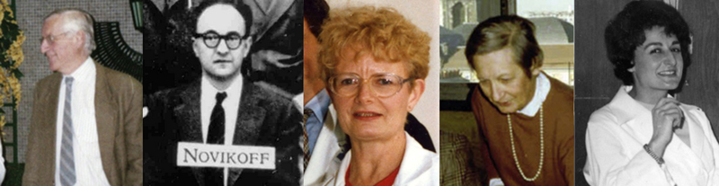
Figure 1: From left to right: Dean Pascal Brunet, Alex Novikoff, Jeanne-Marie Boutry, Françoise Cathala in her office at La Salpêtrière, and Miss Besse, influential secretary of Dean Paul Castaigne. How did a young student in neurology fall into research (nerve tissue culture)? In the Neurosurgery Department of Professor Marcel David, a neurologist, Dr. Elie Bernard-Weil told me one day (yet another stroke of luck!): “I've invented a treatment that allows damaged nerve tissue to regenerate. I'm looking for someone to study it. Would you like to do it on nerve tissue cultures in a research laboratory: the Service de Physiopathologie Cellulaire (Department of Cellular Physiopathology) of Saint-Antoine hospital in Paris?” Of course! It was a turning point. In Doctor Roger Robineaux's laboratory (part of the new research association “Association Claude Bernard pour la Recherche Médicale”), I was given an exceptionally warm welcome by all the members of the teams, including a researcher from the Centre National de la Recherche Scientifique (CNRS), Pierre Goube de Laforest. I learned from them a wide range of tissue culture techniques (of cells: "histiotypic", of organs with migration of certain cells around the explant: "organized", of organs without this cell migration: "organotypic", now called "organoids”) and first applied them to nerve tissue from chicken embryos (one of the least expensive to obtain). I also learned how to study them "in vitro" by phase contrast (or Nomarski) differential interference contrast (DIC) and microcinema or by light and electron microscopy [9]. This made it possible to study both nerve growth factors and the toxicity of certain molecules (e.g., lipidosis induced by some drugs) [10]. I used to go there every afternoon, except for those devoted to internship boarding duty, to the psychology courses I followed, and to the histology tutorials I was giving to young students at the Paris Faculty of Medicine under the supervision of Professor Christian Da Lage for financial reasons (which turned out to be another stroke of luck!). With the help of two women researchers, who were culturing kidneys and livers from human stem cells derived from aborted fetuses, I was able to grow human cerebellar tissue [11]. Ethical issues were quite different in the 1960s! When I asked Pierre Goube de Laforest about human fetal tissue culture, he replied: "I don't see any problem", and, the following week: "I asked around: should we not baptize the fetus before harvesting it, and culturing its nervous tissue?" Among the Doctor Roger Robineaux's laboratory's researchers, two Americans on sabbatical became interested in my nerve tissue cultures. They were Alex Novikoff (Fig 1) and his wife, Phyllis. They used electron microscopy to study lysosomes, recently described by Christian de Duve. They studied the relationship between the neuronal Golgi apparatus and lysosomes. They were helped by an exceptional technician, Jeanne-Marie Boutry (Fig 1), whom they later brought to New York for a year [12-13]. A few years later, they invited me to New York, where I met the American neuropathologist Robert Terry [14] at the Albert Einstein College of Medicine, solidifying my orientation towards neuropathology. How did a young student in neurology fall into neuropathology? A few months later, I met another researcher in Roger Robineaux's laboratory, Françoise Cathala (another stroke of luck!) (Fig 1). She was an exceptional woman - an unconventional neurologist and virologist, very passionate, brilliant, and enthusiastic, but often little understood (even despised) by her colleagues. Clearly interested in my nerve tissue cultures, she whispered to me: "Did you know that an internship position in neuropathology has just been created at La Salpêtrière Hospital in Professor Raymond Escourolle’s new Neuropathology Department? It is alongside Professor Castaigne’s department, just above my virology laboratory. You would be able to perform your nerve tissue cultures there. As no one is yet aware of this new position, you have a great chance of getting the job." Françoise Cathala was right: I realized my dream of becoming an intern at La Salpêtrière. And that's where I met the world of neuropathology with which I was completely unfamiliar, as my training had only included internal medicine, neurology, psychiatry, surgery, and neurosurgery. Neuropathology at La Salpêtrière and La Pitié Hospitals (Paris) in the late 1960s The situation of neuropathology wasn’t simple. The adjacent hospitals La Pitié and La Salpêtrière were still distinct. Initially, there were four different small laboratories ("laboratoires de service") attached to clinical departments [6]. Two were part of the Neurology Departments. The Charles Foix laboratory was headed by Raymond Escourolle (Fig 2, 4 and 6) and belonged to the “Clinique du Système nerveux” headed by the Dean Paul Castaigne (Fig 2) and his very influential secretary, Miss Besse (Fig 1) [4]. His numerous assistants included Pascal Brunet (another lucky break: Miss Besse appreciated Pascal Brunet and helped me in any way she could). When I joined as a resident, this laboratory also carried out neuropathology procedures for the neurological clinics run by Professors André Buge and François Lhermitte. The other neuropathology laboratory of the Neurology Department was located at the “Clinique neurologique” of La Salpêtrière and was headed by Raymond Garcin, followed by Georges Boudin (managed by Michel Fardeau, Jean Lapresle, Jacqueline Mikol, and others) [5,15]. The two other neuropathology laboratories were part of the Neurosurgery Department. One was located at La Salpêtrière (in the department of Professor Jacques Lebeau, successor to Jean-Marie Guillaume who established it in 1942, with the neuropathologist Jean-François Foncin, successor to Jean-Emmanuel Grüner). The other was located at La Pitié Hospital in the Neurosurgery Department of Professors Marcel David (who was followed by Bernard Pertuiset and Jacques Philippon). The neuropathologists of this department were histologists Roger Messimy, Christian Da Lage, Jean Racadot, Jacques Poirier and Michèle Kujas [5-6]. In addition to these neuropathology laboratories, there was also a General Pathology Department (headed by Guy Chomette, then Yves Le Charpentier and Isabelle Brocheriou). This last one used to carry out all the general pathology activity, including autopsies, for patients from non-neurological departments.
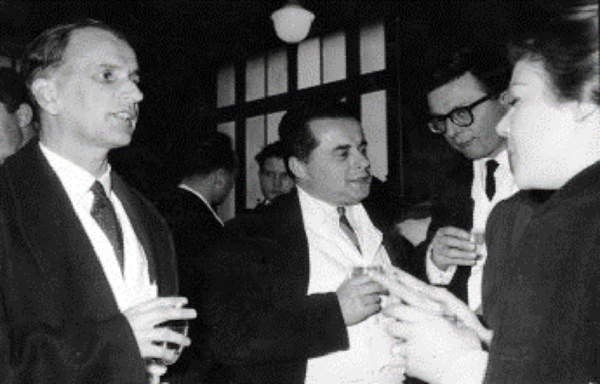
Figure 2: From left to right: Dean Paul Castaigne, Raymond Escourolle and Jean-Claude Gautier. Then, gradually, the various neuropathology "laboratoires de service" were grouped together in the Charles Foix (currently known as Raymond Escourolle) Neuropathology Department. This one progressively took over all the autopsies of the Pitié-Salpêtrière Hospital, and then those of the Parisian region (whether or not they concerned patients with neurological or neurosurgical conditions). This was not without some adventures. The most amusing was the retirement of Jean-François Foncin, which led to the closure of his laboratory. This quirky neuropathologist had decided to take home (in the Paris suburbs) a number of brain samples! But the many formalin-fixed brains that remained had to be moved from the cellar below his laboratory. Together with the Administrative Director of the Pitié-Salpêtrière Hospital Group, we decided to organize their transfer to the Raymond Escourolle Neuropathology Department. To ensure that the operation did not shock hospital staff and patients, the Director staged "renovation work" on J-F Foncin's laboratory. Then "Monsieur Grimi" (Abdelmalek Grimi, technician specializing in autopsies) and I discreetly transported these brains and the related files in my car over several trips. Today, the remaining facilities are the General Pathology Department and the Raymond Escourolle Department of Neuropathology with one exception. Certain muscle biopsies are carried out at the "Institute of Myology", set up in 1996 at the instigation of an association of patients with muscular diseases and their parents, the AFM-Téléthon. Michel Fardeau and his students launched this activity. Charles Foix Department of Neuropathology in the late 1960s The Charles Foix Neuropathology Department carried out cerebrospinal Fluid (CSF) cytology examination and muscle and nerve biopsies (with a dedicated sampling room, where we performed them). Above all, autopsies were by far the most important activity for the intern. I used to perform 4 to 10 per week, first under the guidance of Professor Raymond Escourolle with the help of “Monsieur Grimi”, then alone with him. Later, when I was an intern in clinical neurology at La Salpêtrière, I was sometimes asked by the manager of the neuropathology laboratory to help her intern carry out autopsies. These led to brain-cutting sessions and clinicopathological correlations for multiple departments at La Salpêtrière, but also for other hospitals, such as the Hospice d'Ivry sur Seine (renamed Hôpital Charles-Foix in 1976) or Saint-Germain-en-Laye hospital. Raymond Escourolle (1924-1984), who directed the Charles Foix Neuropathology Department, was a pupil of Paul Castaigne (Fig 2) initially trained in clinical neurology and then mentored in neuropathology by Serge Brion. He succeeded Yvan Bertrand whose predecessors had been Jean-Martin Charcot, Pierre Marie, and Charles Foix. When I arrived in this laboratory, Raymond Escourolle was surrounded by numerous collaborators, all former neurologists: Brigitte Berger, an electron microscopy specialist, Gérard Percheron (Fig 3), an anatomist who had described a variant of the human posterior cerebral circulation (a solitary arterial trunk supplying blood to the paramedian thalami and the rostral midbrain bilaterally) and Jacques Poirier (Fig 3), a histologist and specialist of medicine history. There were also a number of clinicians who performed specialized clinico-pathological research, such as Christian Derouesné, Jean-Claude Gautier (Fig 2), Jean-Louis Ribadeau-Dumas and Jean-Louis Signoret (Fig 3). When Raymond Escourolle died [16], I became head of the Neuropathology Department (which took his name), and was followed by Charles Duyckaerts (Fig 3 and 7), and Danielle Seilhean (Fig 7).
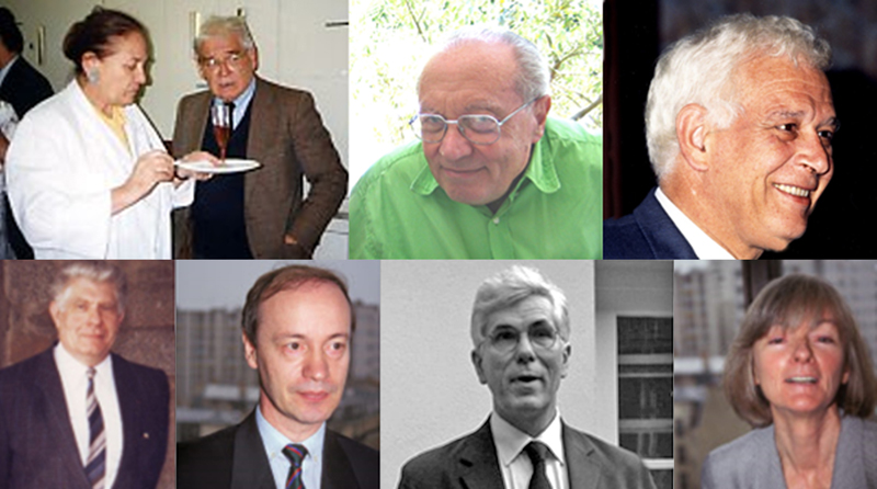
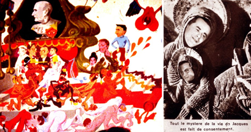
Figure 3: From left to right. Above: Nicole Baumann with Gérard Percheron; Jacques Poirier; Jean-Louis Signoret. Below: General Louis Court; Dominique Dormont; Charles Duyckaerts and Annick Alpérovitch. An internship at La Salpêtrière: the guardroom The residents (“internes”) had some advantages, the most significant of which was the use of the guardroom (“Salle de Garde”). They slept there when they were on night duty and, most importantly, had lunch there whenever they wanted. “Frescoes" decorated the guardroom. These were murals, mostly erotic cartoons, depicting the hospital doctors (Fig 4-6) [17]. After a few introductory rites, the “Salle de Garde” was an opportunity to meet the other interns, the foreign trainees and also the so-called "fossils" that became “dinosaurs” when older. These were the former residents who continued to frequent the guardroom and performed the rituals there ("welcoming newcomers", "bawdy guardroom songs”, etc.). One of the most addicted to the Salle de Garde was the neuropathologist Jean-François Foncin, whom I met there before becoming a fossil myself. He was a remarkable singer, a great specialist in "Chansons de salle de Garde" (guardroom songs), which are usually erotic and divisive.
Figure 4: Raymond Escourolle at the Salpêtrière guardroom with mural “frescoes” circa 1960.
Figure 5: Mural “frescoes” at the Salpêtrière guardroom circa 1980. Bottom right: "The whole mystery of Jacques' life is made of consent".
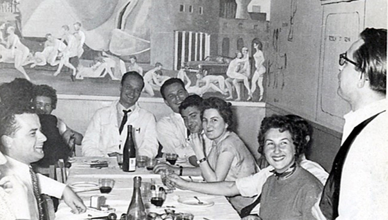
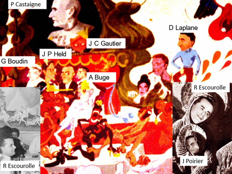
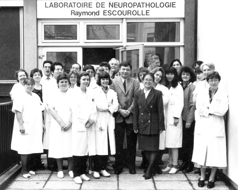
Figure 6: The 1980 “frescoes” of the Salpêtrière guardroom with the names of the people caricatured. The arrival of prion diseases in France Françoise Cathala, who had just returned from the laboratory of future Nobel Prize winner Carleton Gajdusek's in Bethesda, was unique among researchers in France in that she was in love with latent viruses and prion diseases. "I was fascinated by the notion of a latent virus in the nervous system after a primary infection with occasional manifestations appearing subsequently, a phenomenon well known with herpes simplex virus and the herpes virus of varicella–zoster, the subject of my thesis. I wanted to research slow virus diseases with the intention of eventually studying multiple sclerosis, then without any knowledge regarding kuru or the transmission of Creutzfeldt–Jakob disease to chimpanzees!" [18] This caused her some career difficulties (notably at the French research Institute INSERM), as these unconventional infectious agents were far from unanimously accepted by the infectiologists [19]. Actually, the transmission of bovine spongiform encephalopathy from cattle to man had not yet been recognized at that time. It was thanks to her that I met the French pioneers in this field, the researcher Dominique Dormont (Fig 3) and the army medical officer General Louis Court (Fig 3). Together, we carried out the first French autopsy on a chimpanzee inoculated with Creutzfeldt-Jakob disease at the Percy Armed Forces Training Hospital. I took part in numerous activities, including the "Comité sur les encéphalopathies subaiguës spongiformes transmissibles et les Prions” (Committee on transmissible spongiform encephalopathies and prions) headed by Dominique Dormont and known as the "Dormont Committee", and in research and meetings on the subject in collaboration with Jean-Philippe Brandel, Jean-Philippe Deslys, Stéphane Haïk and Corinne Lasmézas. These activities resulted in the establishment of the neuropathological diagnostic criteria for prion diseases [20] and the discovery of the phenomenon of "transconformation", or the spread of protein misfolding from cell to cell. This was a new paradigm in the field of neurodegenerative disorders, brain aging and several other disorders [21]. A passion: research After being introduced to research through nerve tissue culture, and being made aware of prions, I aimed to perform and encourage research practical for the field of neuropathology and, more broadly, for clinical, neurological or neurosurgical disciplines. I carried out this project under the direction and with the help of the INSERM neurochemistry researcher, Nicole Baumann (Fig 1), and then with the epidemiology researcher Annick Alpérovitch (Fig 3). One of my main themes was the neuropathology of dementia (particularly degenerative diseases: Alzheimer's, Parkinson's, Steele-Richardson-Olszewski, fronto-temporal, vascular and 'mixed' dementias as well as the infectious diseases AIDS and prions). I initiated prospective clinical, genetic, biochemical and pathological studies related to this theme with several clinical teams from the Pitié-Salpêtrière (notably the departments of Professors Yves Agid and Olivier Lyon-Caen, François Bricaire and Marc Gentilini), Charles Foix Hospital and other long-stay centers. For instance, we investigated statistical links between the density and distribution of cerebral lesions and the presence, type and intensity of cognitive disorders. Other studies aimed to provide new insights into the neuropathology of Alzheimer's disease, mixed dementia and AIDS. Development of research techniques in the neuropathology department As soon as I was appointed “Assistant” at the Neuropathology Laboratory of the Pitié-Salpêtrière Hospital, I was determined to develop research techniques of the nervous system. In addition to electron microscopy and histochemistry, we introduced tissue cultures, morphometry, immunohistochemistry and molecular biology (in situ hybridization, PCR, nested PCR, in situ PCR). A confocal microscope was installed (Institut Fédératif de Neurosciences de La Salpêtrière). A BSL3 laboratory (P3) was built on the premises of the Pitié-Salpêtrière Faculty of Medicine. It enabled the study of diseases that may be caused by prions and other infectious agents under safety conditions. Neuropathology and Autopsy Four main varieties of autopsies are distinguished in France: the forensic autopsy, medical scientific autopsies, the “gift of corpse to the University” for teaching, and the sanitary autopsy. The Neuropathology Department at La Pitié-Salpêtrière carried out the largest number of medical-scientific autopsies in France. Unfortunately, the number of autopsies requested has fallen steadily since diagnostic techniques, particularly radiological, have improved. This decline is of concern for three reasons. For one, the predictive value of clinical diagnosis (even after using the most modern diagnostic methods) is still poor in some cases, especially when several conditions co-exist. The proportion of unexpected findings that may have modified the patient therapy (20-25 %) has remained unchanged for many years. Second, the autopsy is an important piece of the health watch, i.e., of public welfare. Third, modern post-genomic research needs tissue samples that often cannot be obtained by other means for ethical reasons. I have also carried out neuropathological examinations of forensic autopsies performed by Dominique Lecomte at the Paris Institute of Forensic Medicine. Numerous patients’ associations campaign for making gifts of organs for research purposes easier. We stress the importance of autopsy among medical doctors and caregivers, the prerequisites for restarting autopsy activity and the modifications of regulations and practices that are required [22-23]. Neuropathology of stroke At the Charles Foix Laboratory, autopsies were carried out according to a very strict protocol. In addition to the classic autopsy and the removal of the brain and the spinal cord, a special technique was used to allow a complete examination of the cerebral circulation. Brought back from England by Professor Jean-Claude Gautier, it consisted of whole block removal of the aorta and vessels of the neck, cervical spine, and skull base, including the courses of the carotid and vertebral arteries and the cerebral venous circuits. The block was dissected after formalin fixation, and the results of the macroscopic (and, if necessary, microscopic) examination were recorded on pre-printed diagrams. This allowed very detailed studies of the lesions of the vessels destined for the brain, giving at times surprising results [24-25]. A young neuropathologist's career After my residency, I was successively “Assistant”, “Chef de travaux-assistant”, “Professeur agrégé”, Professor of Pathology and Laboratory Head”, then “Consultant” and “Professor emeritus” at La Salpêtrière. This enabled me to become a member of the "Club français de neuropathologie”, which became the “Société française de neuropathologie." I will always remember the meetings of this society, particularly the one held in Marseille at the invitation of Dean Maurice Toga, when I went in the car of Raymond Escourolle. I was surprised and delighted to discover, in addition to the interesting scientific papers, the famous Marseilles creeks from a sailboat Maurice Toga made available to his guests! I also remember my (unplanned) participation in the 6th International Congress of Neuropathology which was held in Paris (1970), where I discovered with admiration Magdeleine Bérard-Badier, Serge Brion, Edith Farkas, Jean-Emmanuel Gruner, Gilles Lyon, Ludo van Bogaert, Julio Oscar Trelles and many other international leading neuropathologists. I then became a member (and for a time Vice-President) of the European Confederation of Neuropathological Societies [26]. The Charles Foix, then Raymond Escourolle Laboratory: a second family The daily stays in the laboratory, the professional and often friendly contacts with all the members of the laboratory (head of department, colleagues, assistants, interns, secretaries, technicians, nurses (Fig 7)), the biopsy, cytology and autopsy procedures ... quickly led to what might be considered an addiction! Working in the laboratory wasn't just a duty or a routine. It was often a pleasure. All the more so because some of the events that took place were puzzling (and sometimes very amusing!).
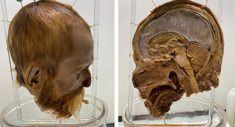
Figure 7: Group photo of members of the Raymond Escourolle Laboratory in 1994. An exceptional story: Ravachol’s head François-Claudius Koënigstein (more commonly known as Ravachol) was a French anarchist militant, born on October 14, 1859, in Saint-Chamond (between Saint-Etienne and Lyon in France). Having committed several offenses, murders, and attacks, he was guillotined in Montbrison, near Saint-Chamond, on July 11, 1892. As was usual at the time, half his head was preserved in a glass jar containing formalin, and given to “Science”, i.e. in practice, to Jean-Martin Charcot’s laboratory. When I was resident, Ravachol's head was kept in Professor Escourolle's office, in a locked cupboard with a glass door, allowing it to be admired (Fig 8). A few years later, when I arrived in the Neuropathology Department in April 1975, police special forces occupied the site as the head of Ravachol had been stolen! Indeed, Raymond Escourolle's wardrobe had been broken into and the head had disappeared. Within a few days, I understood what had happened from the gossip in the on-call room. Some inebriated residents (one of whom worked in André Buge’s department) had stolen the head as a gift for one of their girlfriends (granddaughter of Karl Marx, who cried out in horror at the sight of this gift!). Not knowing what to do, the friends came up with a fantastic idea to make a splash. And this was implemented a few days later: Ravachol’s head was found in front of the Pantheon, a mausoleum for the remains of distinguished French citizens in the 5th arrondissement of Paris, modelled on the Pantheon in Rome. It was accompanied by a missive: « Ceci est la tête de Ravachol. Elle devrait être au Panthéon, Et non pas chez Escourolle. » (“This is Ravachol’s head. It should be in the Pantheon, not at Escourolle’s”). And the story was confirmed by the concerned intern Jean-Christophe Rufin, who gave up medicine, became a humanitarian (notably in Ethiopia), then French ambassador, then novelist and member of the renowned Académie Française who later recounted it in his book: “Un Léopard sur le garrot” [27].
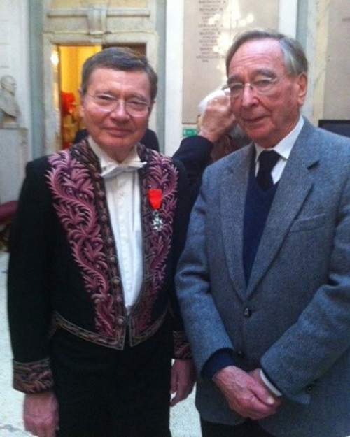
Figure 8: Ravachol’s head (Sorbonne University Collection, under the inventory number: SU.PM.T.2023.0.1.). Administrative work and learned societies Other strokes of luck also befell me! Very early on, I was appointed Director of the “Experimental and Clinical Neuropathology” associated training course, then of the “Neurology, Neuropathology” Center of the Claude Bernard Association. I was elected as a member of the Board of Directors of this Association (later renamed the Robert Debré Association for Medical Research). I was appointed treasurer, a post I still hold. I was then elected a member of the Medical Committee of Pitié-Salpêtrière Hospital, then Representative of the Pathologists on the Medical Committee of Assistance Publique des Hôpitaux de Paris. I was then appointed a member of the Pathological Anatomy Section of the French Conseil Supérieur des Universités. Professor (now Dean) Pascal Brunet asked me to become a member of the Board of Directors of the Pitié-Salpêtrière Faculty (I have also been Vice-Dean). I was also appointed a member of the Institut Fédératif de Neurosciences de La Salpêtrière and Vice Chairman of the Specialized Scientific Commission 6, Neurosciences B of the French national medical research institute (INSERM). Then, for obscure reasons (probably to avoid the election of someone else), I was asked by Jean Cambier and Jean-Claude Gautier to apply for membership of the French National Academy of Medicine, which had never had a neuropathologist member before. And for the same obscure reasons, with the help of pathologist Louis Orcel, I was elected (by just one vote!). This gave me the opportunity to rub shoulders with the leading French and foreign specialists in all areas of human and animal medicine and pharmaceuticals and to help draft numerous press releases and reports for the French authorities. I was later elected Treasurer for four years (Fig 9) and remain Chairman of the "Mental Health, Neuroscience and Addiction" Commission. I was also given the opportunity to develop, with Professor Jean-Paul Tillement, the French Academy of Medicine's Franco-Québec relations.
Figure 9: Dressed as an Academician, when I was treasurer with Professor Jean Cambier. A large number of countries visited and host of colleagues and friends from all over the world I was fortunate enough to be invited to give conferences in most French regions (including Guadeloupe) and many other countries (including Argentina, Australia, Austria, Belgium, Canada, Germany, Hungary, Israel, Italy, Japan, Peru, Spain, Sweden, Tunisia, the United Kingdom and the United States of America (USA)), which gave me the opportunity to make some very interesting visits. I have always been warmly welcomed, and have also had the pleasure of welcoming and working with a large number of foreign colleagues, some of whom came to live in France (in particular from Algeria, Armenia, Australia, Belgium, Brazil, Canada, China, Colombia, Germany, Hungary, India, Italy, Iran, Lebanon, Madagascar, Mexico, Morocco, Japan, Peru, Poland, Portugal, Romania, Russia, Spain, Switzerland, Tunisia, United Kingdom and the USA (Fig 7)). I remember the friendly welcomes I received in many countries, notably Germany, Italy, and Spain. The relationships that gave me the best memories are those developed with five countries: Peru, India, USA, Japan, and the United Kingdom. In Peru, thanks to the invitation and warm welcome of Julio Oscar Trelles [28], a former student of Jean Lhermitte in Paris (and his son Luis, who entered the Charles Foix Laboratory), it was a delight to discover one of the wonders of the world: Machu Pichu. It was also a pleasure to welcome T. Vasudev Rao, a pathologist from Bangalore. During our holidays we were able to lend him the Paris flat we were renting, overlooking the Seine at the foot of the Eiffel Tower, which enabled him to spend a few days there with his family. In gratitude, he gave us a very pretty Hindu statue, which still hangs in our flat in Dole. Umberto de Girolami, a neuropathologist at Brighman and Women’s Hospital in Boston (USA), stayed several times at La Salpêtrière and returned regularly to Paris as a foreign member of the French Académie de Médecine. As far as Japan is concerned, I had the pleasure of discovering Kyoto, Nara and Tokyo with Toshiki Uchihara. In the United Kingdom, it was Queen’s Square National Hospital and the Institute of Neurology in London. Professors Leo Wilfried Duchen and Francisco Scaravilli invited me, and came to Paris in return, enabling us to make fruitful contacts and even enjoy pleasant trips several years later. It also gave me the opportunity to meet my second wife, Chantal Hausser, a Canadian neurologist, and neurophysiologist who had been a trainee for 3 months at the Charles Foix Laboratory and whom I met again 12 years later in Montreal. I had the pleasure of welcoming some of these foreign colleagues (Leo Duchen, Umberto de Girolami and Toshiki Uchihara) in our house in Dole or in our country house at the Abbey of Baume les Messieurs, in the French Jura, and taking them on a tour of the landscapes and little-known beauties of this area (Dole and Arbois, the cities of Louis Pasteur, the Cirque de Baume-les-Messieurs and Chateau-Chalon, the Royal saltworks of Arc et Senans, lake Chalain, the medieval city of Nozeroy etc.). A difficult (and risky) diagnosis thanks to my English friends! One of my colleagues and friends at La Salpêtrière told me, "I've just admitted to Neurosurgery one of my patients, the wife of a very important person. She has a brain tumor. Can you let me know what the diagnosis is?” I couldn't forget his request. A few days later, I received a message from a pathologist in the United States asking me, on behalf of the VIP, for some slides from this biopsy. I sent them with the agreement of the attending physician. The diagnosis seemed obvious to me but, consulting recent publications on the subject, I also decided to send some white slides to Francisco Scaravilli. What a surprise it was to receive, a few days later, an invitation to meet a North American delegation in Paris! In addition to the American pathologist, the meeting included a Quebec pathologist who was supposed to translate English into French, and a series of secretaries and other people whose role was unclear to me. It was decided to hold two "multidisciplinary meetings" first with microscopes, at the Raymond Escourolle Laboratory, then at the department of my colleague, the patient’s attending physician. Thanks to the notoriety of Francisco Scaravilli, I could impose the diagnosis, and only after the opinion of another colleague, a hematologist and oncologist at La Salpêtrière, could we start the treatment best suited to the patient’s illness! The new developments in neuropathology Whatever the procedure - brain biopsy, now performed very often by stereotaxy [29], including very challenging area such as the brain stem [30], muscle and nerve biopsies, skin biopsies for quantifying small nerve fibers, cytological examination of cerebrospinal fluid or autopsy - neuropathology techniques have recently exploded and are continuing to develop. For example, stimulated Raman scattering microscopy, a label-free optical imaging method performed on smear or frozen-sections, is developing to quickly generate digital hematoxylin-and-eosin-stained-like images for histopathological tissue analysis [31]. It can be used in the operating room to provide fast answers for brain biopsies. Of course, artificial intelligence also enters the world of neuropathology. Curtis Langlotz’s, Director of the Center for Artificial Intelligence in Medicine and Imaging of Sanford University, formula about radiologists [32] can be stated as: “Artificial intelligence won't replace neuropathologists, but neuropathologists who use artificial intelligence will replace those who don't!” Why fall into neuropathology? Firstly, it is sometimes a matter of chance, which I would not hesitate to describe as fortunate. Secondly, it has many advantages. Some of these are very material: for example, it is an exception for a neuropathologist to work on weekends (apart from writing scientific articles or preparing lectures or presentations for scientific conferences) or to be summoned in a hurry! Other advantages are more intellectual in nature. They involve, for example, puzzle solving: the cause(s) of any nervous system illnesses the patient may have or have had, or establishing the precise nature of a tumor and its prognosis, using increasingly sophisticated techniques, to which we must constantly adapt. To continue the comparison with games, it's the equivalent of a chess game or bridge, played with increasingly strong partners. Artificial intelligence, far from being able to replace the experience and knowledge of the neuropathologist and his numerous coworkers, must now be used as an additional tool. And this requires a great deal of companionship, which is always important in medicine, but even more so in neuropathology, where getting things wrong can have serious consequences. So, neuropathology requires the development of multiple skills in a wide range of clinical and biological disciplines, from pathology and neurology to genetic and molecular biology. It therefore imposes a wide range of complementary training, which often proves fascinating. In addition, this discipline, more than most other medical ones, offers a wide variety of diagnostic, teaching, and research activities. What's more, this rare specialty brings us into contact with many colleagues from different regions and countries, leading to numerous trips and the discovery of other points of view and even civilisations. Last but not least, to sum up: it's a fascinating job! By way of conclusion, taking inspiration from Rudyard Kipling: « If you can dream – and not make dreams your master; If you can think – and not make thoughts your aim; Or walk with Kings – nor lose the common touch, If neither foes nor loving friends can hurt you, If all men count with you, but none too much; And – which is more – you’ll be a Neuropathologist, my son! » Acknowledgements I would like to thank Danielle Seilhean, who provided Figures 7 and 8, and Chantal Hausser-Hauw, who reviewed this article. References 1. Gandolfo G, Deschaux O (2010) Histoire de la découverte du cerveau et de l’évolution des méthodes d’exploration: de la Préhistoire à nos jours [History of the discovery of the brain and the evolution of exploration methods; from prehistory to today]. Biologie Géologie 2-2010, pp.127 hal-01090539. 2. Paillette C, Griset P, Agid Y (2021) La genèse des neurosciences - Entre technosciences et diplomatie de l’innovation, des années 1940 aux années 1970 [The genesis of neurosciences: Between technosciences and innovation diplomacy, from the 1940s to the 1970s]. Med Sci (Paris) 37(10):920-926. https://doi.org/10.1051/medsci/2021139 3. Bonduelle M (1997) La Salpêtrière de Mazarin à Charcot. French [The Salpêtrière from Mazarin to Charcot] Hist Sci Med 31(2):163-170. French. PMID: 11625157. 4. Cambier J (1997) Histoire de la Salpêtrière: Alajouanine puis Castaigne. L'ouverture. French [History of La Salpêtrière: Alajouanine then Castaigne. The opening]. Hist Sci Med 31(2):201-4. French. PMID: 11625162. 5. Walusinski O, Poirier J (2017) L’essor de la neuropathologie au service de la clinique à La Salpêtrière (1862-1923) [The development of neuropathology at the service of the clinic at La Salpêtrière (1862-1923)]. In « Le cerveau au microscope: la neuroanatomie française aux XIXe et XXe siècles » [The brain under the microscope: French neuroanatomy in the 19th and 20th centuries] J.G. Barbara, F. Clarac (eds), Paris, Hermann. 6. Seilhean D (2019) Neuropathology in Pitié-Salpêtrière Hospital: Past, present and prospect. Neuropathology 40(1): 3-13. https://doi.org/10.1111/neup.12616 7. Poirier J (2012) L’externat des hôpitaux de Paris (1802-1968) [Paris hospital externship (1802-1968]. Collection Histoire des sciences, Paris, Hermann, 394 p. ISBN: 978-2-7056-8426-6. 8. Philippon J (1997) Histoire de la Neurochirurgie à la Pitié Salpêtrière [History of Neurosurgery in la Pitié-Salpêtrière]. Hist Sci Med 31:173-180. French. PMID: 11625159. 9. Hauw JJ (1968) Etude morpho-dynamique et ultrastructurale du ganglion spinal d’embryon de poulet en cultures organisées ou organotypiques. Thèse de Doctorat en médecine [Morpho-dynamic and ultrastructural study of the chicken embryo spinal ganglion in organized or organotypic cultures. Thesis for Doctorate in Medicine, Paris. 10. Hauw JJ, Boutry JM, Hamam S, Escourolle R (1978) Lipidose médicamenteuse induite en culture de ganglion spinal de Souris par le maléate de perhexiline. Résultats préliminaires concernant la toxicité aiguë du médicament [Perhexiline-maleate-induced lipidosis in mouse spinal ganglia tissue culture. Preliminary results on the acute toxicity on the drug]. C R Acad Hebd Seances Acad Sci D 287(10):959-61. French. PMID: 106980. 11. Hauw JJ, Berger B, Escourolle R (1972) Présence de synapses en culture organotypique in vitro de cervelet humain [Presence of synapses in organotypic culture in vitro of human cerebellum]. C R Acad Hebd Seances Acad Sci D 274(2):264-6. French. PMID: 4622074. 12. Hauw JJ, Novikoff AB, Novikoff PM, Boutry JM, Robineaux R (1972) Culture of nervous tissue on collagen in Leighton tubes. Brain Res 37(2):301-9. https://doi.org/10.1016/0006-8993(72)90675-0 13. Novikoff PM, Novikoff AB, Quintana N, Hauw JJ (1971) Golgi apparatus, GERL, and lysosomes of neurons in rat dorsal root ganglia, studied by thick section and thin section cytochemistry. J Cell Biol 50(3):859-86. https://doi.org/10.1083/jcb.50.3.859 14. Terry N, Masliah A, Overk C, Masliah E (2019) Remembering Robert D. Terry at a time of change in the world of Alzheimer's disease. J Alzheimers Dis 70(3):621-628. https://doi.org/10.3233/JAD-190518 15. Mikol J (2021) Notes on the career of Jacqueline Mikol. Free Neuropathology 2:25. https://doi.org/10.17879/freeneuropathology-2021-3532 16. Hauw JJ (1984) In memoriam Dr. Raymond Escourolle (1924-1984). Acta Neuropathol 65:89. https://doi.org/10.1007/BF00690461 17. Godeau E (2009) Les fresques de salle de garde [The guardroom frescoes]. Sociétés & Représentations 2(28):13-30. 18. Cathala F (2008) Why I joined the research laboratory of Prof. D. Carleton Gajdusek in 1968. Philos Trans R Soc Lond B Biol Sci 363(1510): 3631–3632. https://doi.org/10.1098/rstb.2008.4008 19. Court L, Hauw JJ (2015) Le docteur Françoise Cathala Pagesy et l'histoire des maladies à prions [Doctor Francoise Cathala and history of prions diseases]. Rev Neurol (Paris) 171(12):805-11. https://doi.org/10.1016/j.neurol.2014.02.003 20. Budka H, Aguzzi A, Brown P, Brucher J-M, Bugiani O, Gullotta F, Haltia M, Hauw J-J, Ironside JW, Jellinger K, Kretzschmar HA, Lantos PL, Masullo C, Schlote W, Tateishi J, Weller RO (1995) Neuropathological diagnostic criteria for Creutzfeldt-Jakob disease (CJD) and other human spongiform encephalopathies (prion diseases). Brain Pathol 5(4):459-66. https://doi.org/10.1111/j.1750-3639.1995.tb00625.x 21. Hauw JJ, Haïk S, Duyckaerts C (2015) La transconformation protéique, nouveau paradigme en neurologie [Spreading of protein misfolding: A new paradigm in neurology]. Rev Neurol (Paris) 171(12):825-31. French. https://doi.org/10.1016/j.neurol.2015.09.010 22. Hauw JJ (2001) Les différentes variétés d’autopsie. Propositions pour un renouveau de l’autopsie médicale et scientifique. [The different varieties of autopsy. Proposal for a renewed medical and scientific autopsy]. Bull Acad Natl Med 185(5):829-38. French. PMID: 11717841. 23. Lecomte D, Hauw JJ (2015) Les autopsies médico-scientifiques sont indispensables au progrès médical [Medical and scientific autopsies are essential to progress in medicine] Report by the French National Academy of Medicine, Tuesday 7 April 2015. 24. Amarenco P, Hauw JJ, Hénin D, Duyckaerts C, Roullet E, Laplane D, Gautier JC, Lhermitte F, Buge A, Castaigne P (1989) Les infarctus du territoire de l'artère cérébelleuse postéro-inférieure. Etude clinico-pathologique de 28 cas [Cerebellar infarction in the area of the posterior cerebellar artery. Clinicopathology of 28 cases]. Rev Neurol (Paris) 145(4):277-86. French. PMID: 2660219. 25. Klein IF, Labreuche J, Lavallée PC, Mazighi M, Duyckaerts C, Hauw JJ, Amarenco P (2010) Is moderate atherosclerotic stenosis in the middle cerebral artery a cause of or a coincidental finding in ischemic stroke? Cerebrovasc Dis 29(2):140-5. https://doi.org/10.1159/000262310 26. Mikol J, Weller R (2006). Neuropathology in Europe: an overview. Clin Neuropathol 25(1):7-13. PMID: 16465768. 27. Ruffin JC (2008) Un léopard sur le garrot [A léopard on the withers] Gallimard Ed. ISBN 9782070782901. 28. Escalante-Sanchez S (2005) Evocacion de J Oscar Trelles. Rev. de Neuro-Psiquiat. 68:3-4. 29. Bex A, Mathon B (2023) Advances, technological innovations, and future prospects in stereotactic brain biopsies. Neurosurg Rev 46:5 https://doi.org/10.1007/s10143-022-01918-w 30. Jung IH, Chang KW, Park SH, Moon JH, Kim EH, Jung HH, Kang SG, Chang JH, Chang JW, Chang WS (2021) Stereotactic biopsy for adult brainstem lesions: A surgical approach and its diagnostic value according to the 2016 World Health Organization Classification. Cancer Med 10(21): 7514-7524. https://doi.org/10.1002/cam4.4272 31. Francis A, Berry K, Chen Y, Figueroa B, Fu D (2017) Label-free pathology by spectrally sliced femtosecond stimulated Raman scattering (SRS) microscopy. PLoS ONE 12(5): e0178750. https://doi.org/10.1371/journal.pone.0178750 32. Langlost CB (2019) Will Artificial Intelligence Replace Radiologists? Radiol Artif Intell 1(3):e190058. https://doi.org/10.1148/ryai.2019190058
Copyright: © 2023 The author(s). This is an open access article distributed under the terms of the Creative Commons Attribution 4.0 International License (https://creativecommons.org/licenses/by/4.0/), which permits unrestricted use, distribution, and reproduction in any medium, provided the original author and source are credited, a link to the Creative Commons license is provided, and any changes are indicated. The Creative Commons Public Domain Dedication waiver (https://creativecommons.org/publicdomain/zero/1.0/) applies to the data made available in this article, unless otherwise stated. |