|
|
|
Free Neuropathology 4:16 (2023) |
|
Reflections |
|
My recollections of my 50 years as a neuropathologist from a neurologist’s perspective |
|
Yoshio Hashizume |
|
Institute of Neuropathology, Fukushimura Hospital, Noyori-cho, Toyohashi, Aichi, Japan |
|
Corresponding author: |
|
Additional resources and electronic supplementary material: supplementary material |
|
Submitted: 04 September 2023 |
|
Keywords: Neurodegenerative disease, Pathology of dementia, Spinal cord pathology |
|
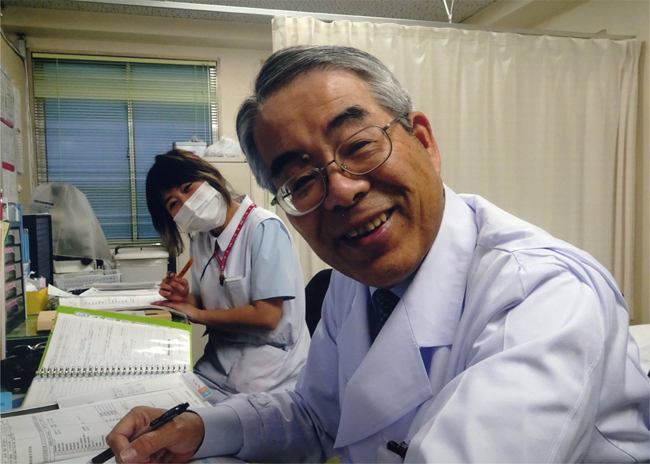
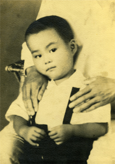
Figure 1: A recent photo of me taken in the outpatient department of Fukushimura Hospital. Nurse Korin Kondo is on the left. Introduction I am very honored to be able to write about my neuropathology background in the Reflections section. Fifty years have passed since I started researching neuropathology, and I am grateful for the good fortune that I have enjoyed to be able to continue working in this field. In this section, I would like to describe the history of my neuropathological work in the Tokai region, which is located in the central part of Japan. Figure 1 is a recent photo of me taken in the outpatient department of Fukushimura Hospital. My background I was born on August 25, 1944 at the end of World War II in the mountain village of Haze in Mie Prefecture, which is located in central Japan. The village of Haze was two hours by car from Matsuzaka, a city in the center of Mie Prefecture, and it was located in a rural area where buses came only three times a day on unpaved roads. Both my parents were from the village of Haze. The village was my hometown. It was where the Kushida River flowed, and it had clean water and beautiful mountains. My father worked as a police officer in the City of Tsu, the prefectural capital of Mie Prefecture. I still vividly remember the city in ruins from when it was destroyed by air raids during the war. I grew up as the second son of four siblings. I was brought up with the immeasurable affection of my parents in a chaotic post-war Japan with scarce supplies. Figure 2 shows a picture of me when I was four years old.
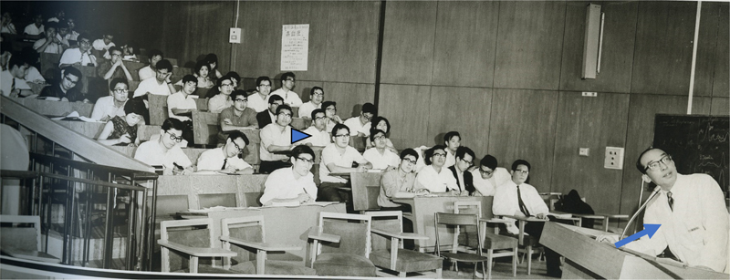
Figure 2: A picture of me when I was four years old. After attending elementary and junior high school in the City of Tsu, I began attending Tsu High School, one of the leading preparatory schools in the prefecture in 1960. Even though my parents weren’t wealthy, they created a warm home and let me study to my heart‘s content. During summer vacation back then, I was studying for 16 hours a day, except for eating and sleeping. When I was in high school, my mother got sick and had to go to the hospital. That‘s why I chose to become a doctor: because I wanted to help people. In 1963, I was admitted to the Nagoya University School of Medicine, which is where I wanted to go. Having grown up in the countryside, Nagoya was a big city for me. I lived in a small boarding house in a room with only 3 tatami mats and no windows. While studying medicine, I was able to lead a fulfilling life with extracurricular activities, student movements, and a part-time job. Figure 3 shows a lecture on neurology by Professor Itsuro Sobue, who was an associate professor at the time. The arrow is to Professor Sobue, and the arrowhead is to me.
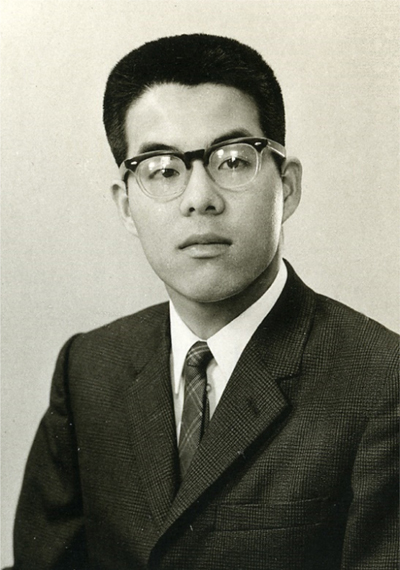
Figure 3: A lecture on neurology by Professor Itsuro Sobue, who was an associate professor at the time. The arrow is Professor Sobue, and the arrowhead is me. After school, I was active in the chorus, and this is where I got to know my wife, who was in the chorus at another university. I was actively involved in political and social issues and participated in student movements. At the time, Japan was in the midst of the Okinawa reversion movement. I wanted to see Okinawa before it was returned to Japan, so I traveled there with a tent. I also worked hard at various part-time jobs, such as selling ice cream and oranges on the platforms of Nagoya Station. Figure 4 is a picture of me when I graduated from Nagoya University School of Medicine.
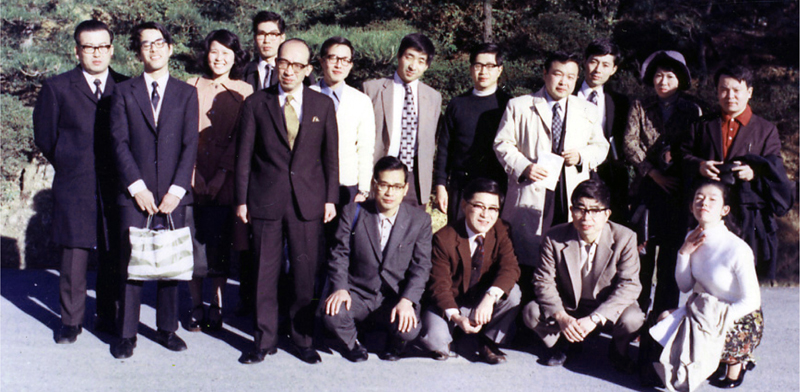
Figure 4: A picture of me when I graduated from Nagoya University School of Medicine. Before I started studying neuropathology After graduating from Nagoya University School of Medicine in 1969, I worked as a physician at Anjo Kosei Hospital in the Mikawa area for two years. This hospital is a leading hospital for acute care in the Mikawa district of Aichi Prefecture, and I was able to diagnose and treat various medical conditions. During these two years, I was involved in pathology autopsies on many patients for whom I was the attending physician. This was a major impetus for me to major in pathology. Around that time, Dr. Izumi Kano, my advisor, told me to “Be the Master of an Art.” When I later majored in neuropathology, I always ruminated on the phrase “Be the Master of an Art” during the procedure to remove the spinal cord at autopsy. I got married while working at Anjo Kosei Hospital and later had a son and two daughters. After practicing general internal medicine for two years, I was clinically involved in neurological diseases in the Neurology Laboratory at the First Department of Internal Medicine, Nagoya University School of Medicine. In the laboratory, I was advised by Professor Itsuro Sobue. Figure 5 is a group photo taken during a trip to Kashikojima with my colleagues from the Department of Neurology, Nagoya University School of Medicine. Dr. Sobue was one of the founders of neurology in Japan, a specialist in peripheral nerve diseases, and he advanced Japanese research in gerontology as a leader in research on geriatric medicine. Dr. Sobue served as president of Aichi Medical University for many years and trained many neurologists in the Nagoya area. The starting point for me in neuropathology was the Department of Neurology, Nagoya University. Clinical experience in neurology was extremely vital to my subsequent interest in neuropathology. At the time, however, there were not enough brains to autopsy in the Nagoya area to study neuropathology. I wanted to search for the brains of patients for whom I was the attending physician and on whom pathological autopsies were performed. Brains for autopsy belonged to the Department of Pathology, and in order to do neuropathology there was no other way than to join the Department of Pathology. Therefore, I joined the Department of Pathology at Nagoya City University in 1974 and began studying systemic pathology under Professor Hidemasa Kishimoto. Dr. Kishimoto specialized in clinical pathology and was of a good character, and many students and clinicians came to study at his laboratory. At the time, my goal was to acquire the skills to perform pathology autopsies by myself even if I was assigned to a local hospital later on. During this period, I trained in basic pathology, including organ excision after fixation, embedding, sectioning, staining, microscopy, summarizing findings, and giving presentations at clinical pathology conference.
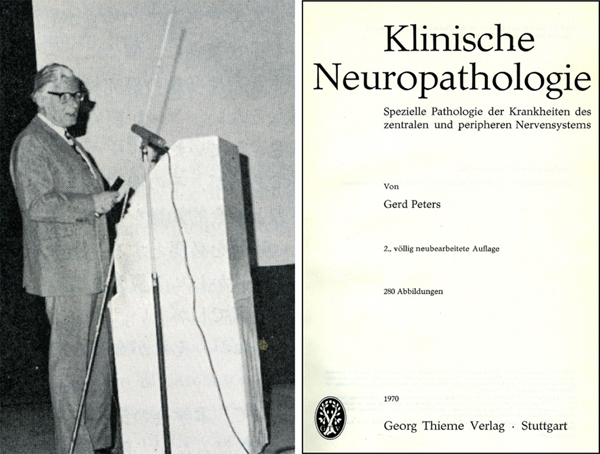
Figure 5: A group photo taken during a trip to Kashikojima with my colleagues from the Department of Neurology at Nagoya University School of Medicine. In the back row, 1st from is Dr Shigemitsu Nishigaki, 2nd is me, 5th is Professor Itsuro Sobue, 1st from the left in the front row is Dr. Akira Takahashi, and 3rd is Dr Tsutomu Yanagi. Studies abroad in Munich In 1974, Dr. Shigemitsu Nishigaki from Nagoya University School of Medicine introduced me to the Max Plank Institute for Psychiatry in Munich, Germany, and I decided to study there. I studied German language for two months at the Goethe Institute in Grafing in the suburbs of Munich before starting my life at the Institute. Learning German with many students from all over the world was a very valuable experience. The Max Plank Institute for Psychiatry is one of Europe‘s leading neuropathology institutes. A professor at the time was Professor Gerd Peters, editor of Acta Neuropathologica and author of Klinische Neuropathologie [Clinical Neuropathology]. This book is an excellent work on neuropathology. On the left in Figure 6 is Professor Peters when he gave a special lecture at the Japanes Society of Neuropathology, and on the right is the textbook Klinische Neuropathologie.
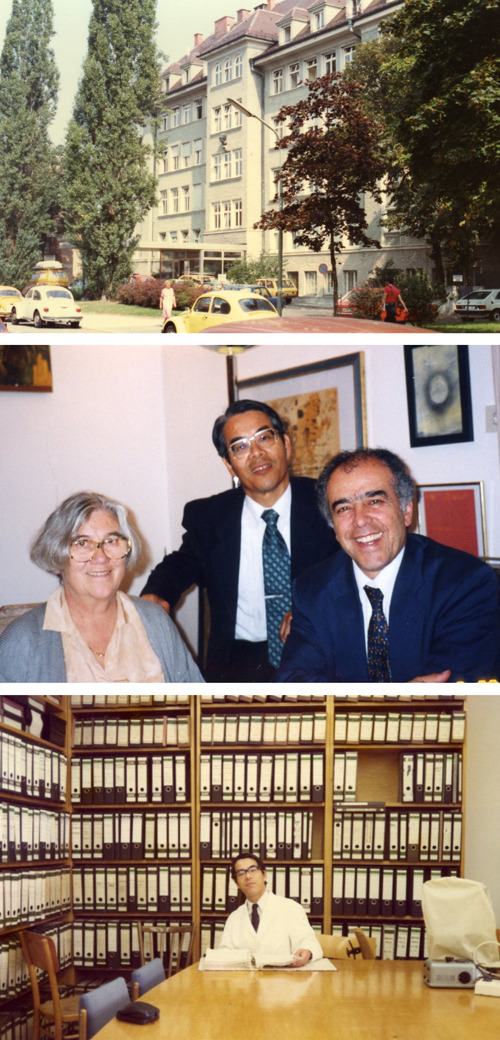
Figure 6: On the left is Professor Gerd Peters when he gave a special lecture at the Japanese Society of Neuropathology. The right is the textbook Klinische Neuropathologie. The Institute received a large number of brains for autopsy from southern Germany, mainly from psychiatric hospitals. I observed Professor Peters cutting the brain and I was able to observe the gross findings from many cases, including multiple sclerosis and Wernicke‘s encephalopathy. I was able to live a blessed life, reading the book Klinische Neuropathologie every day, looking at the pictures in the book, and looking through a microscope every day. Dr. Parviz Mehraein, who was an associate professor in the laboratory at the time, was a boon not only to my research but also to my life in Germany, which I was not accustomed to. The upper of Figure 7 is the Max Plank Institute for Psychiatry in Munich, the middle is Dr. Mehraein, his wife and me, and the lower left is me in the laboratory. Because of this study abroad, I decided to make neuropathology my life‘s work. I was able to publish a paper summarizing the clinicopathological findings of cases of cerebral aqueduct obstruction that were compiled at the Institute (1). In Munich, I was able to study closely with Dr. Motohiro Suetsugu of Kyushu University and Dr. Tatsuji Tanabe of Nihon University, who came from Japan to study abroad.
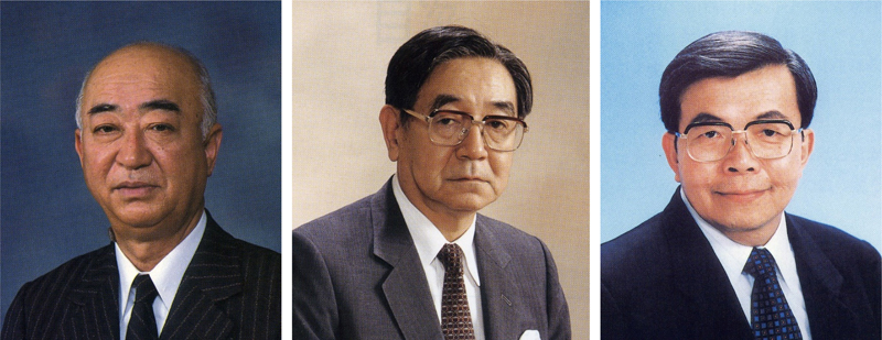
Figure 7: Upper: The Max Plank Institute for Psychiatry in Munich. Middle: Dr. Parviz Mehraein, his wife and me. Lower: me in the lab. Obtaining a Doctorate in Medicine After studying abroad in Munich, I returned to Japan in 1976 and continued my research on neuropathology while continuing to study general pathology under Professor Kishimoto in the Department of Pathology at Nagoya City University. Advised by Dr. Kishimoto and Dr. Tsutomu Yanagi, head of the Department of Neurology, Nagoya Daini Red Cross Hospital, I received a doctorate in medicine for my pathological research on ossification of the posterior longitudinal ligament (2,3). As a neurologist, Dr. Yanagi was an excellent doctor who was familiar with spinal cord diseases such as cervical spondylosis, ligamentous ossification, and spinal cord infarction. After I was advised by Dr. Yanagi, I decided to make spinal cord pathology my life’s work. The research on ossification of the posterior longitudinal ligament was later taken over by Dr. Takashi Kameyama (4) who published a wonderful paper in the journal Brain. Life as a researcher in the Department of Pathology, Nagoya University In 1980, I was hired as an assistant in the Department of Pathology at my alma mater, Nagoya University. I was able to continue my research in neuropathology under Professor Soichi Iijima, who was an instructor in the Japanese Society of Pathology. Dr. Iijima specialized in pathology of the spleen, but he allowed me to continue with neuropathology. He told me that I should devote myself wholeheartedly to neuropathology. He had a wonderful attitude toward pathology autopsies, and he wrote in his book that “Problems that need to be addressed can always be found in the body of a deceased patient, and how to deal with those problems depends on the ability of the clinician and the pathologist.” Taking these words to heart, I have devoted myself to pathology autopsies (5). Later, Dr. Junpei Asai became a professor. Dr. Asai specialized in pathology of the spleen. During his tenure as professor, he was stricken with malignant lymphoma, but he fought the disease with fortitude and completed his professorship. I was indebted to this laboratory for 13 years until I was transferred to Aichi Medical University. Figure 8 shows a photograph of the teachers who taught me pathology. Professor Kishimoto is on the left, Professor Iijima is in the middle, and Professor Asai is on the right.
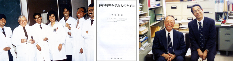
Figure 8: The teachers who taught me pathology. From left, Professor Hidemasa Kishimoto, Professor Soichi Iijima, and Professor Junpei Asai. Study abroad at Montefiore Hospital In 1982, I was a researcher of the Ministry of Education and I studied at Montefiore Hospital in New York City, where I was able to train in neuropathology under Professor Asao Hirano. This was around the time that AIDS broke out in New York, and cases for autopsy began to appear. Being able to study with Dr. Imaharu Nakano and many other doctors from Japan in Professor Hirano’s laboratory was very helpful in continuing my study of neuropathology in Japan. With Professor Hirano’s guidance, I was able to write a paper on spinal cord intramedullary metastasis and spinal pencil-shaped softening (6,7). Dr. Hirano’s lab was visited by many doctors from Japan who wanted to study neuropathology, such as neurologists, neurosurgeons, psychiatrists, and pathologists. Dr. Hirano made a massive contribution to the advancement of neuropathology in Japan. Dr. Hirano’s famous book “For Those Who Study Neuropathology” became a bestseller. In Figure 9, the left is a moment at the laboratory with professors Zimmermann and Hirano and research students from Japan. The center is Professor Hirano’s famous book “For Those Who Study Neuropathology.” The right is Professor Hirano and me in my professor’s office.
Figure 9: The left is a moment at the laboratory with professors Zimmermann and Hirano and research students from Japan. The center is Professor Hirano’s famous book “For Those Who Study Neuropathology.” The right is Professor Hirano and me in my professor’s office. Conducting research in the Pathology Department, Nagoya University Hospital In 1990, I was appointed as an associate professor in the Department of Pathology, Nagoya University Hospital. With the help of pathologists, neurologists, and neurosurgeons from major hospitals in the Tokai region of central Japan, we focused on collecting brains for autopsy. The brain can be searched for many crucial neurological diseases, and clinical neuropathological conferences are conducted at many hospitals. At the time, the director of Nagoya University Hospital was Professor Akira Takahashi, a neurologist whom I respect, and I was able to conduct research along with many young neurologists in his department. Figure 10 is a group photo of me and my colleagues when I was in the Department of Pathology, Nagoya University Hospital. Here are the papers with many doctors who were conducting joint research at the time (8-19).
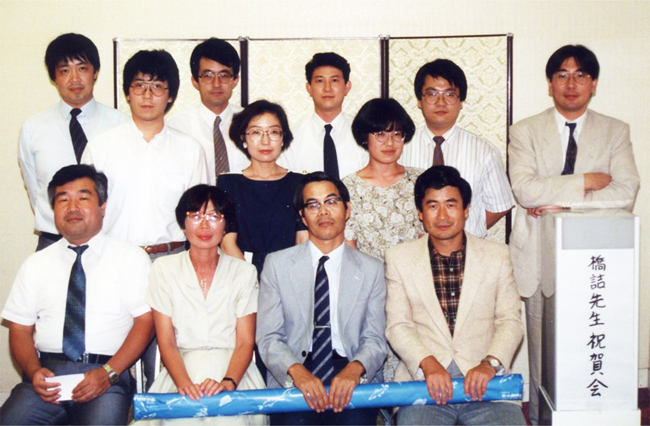
Figure 10: A group photo of me and my colleagues when I was in the Department of Pathology, Nagoya University Hospital. Front row from left: Toshiaki Inagaki, Noriko Hirunagi, me, and Shigeo Riku. Middle row: Mari Yoshida and Motoko Sakai. Back row from left: Tetsuo Ando, Tetsuya Mizuno, Satoshi Okuda, Takashi Kameyama, Shinichi Miyao, and Akito Kume. In western Japan, there is an academic conference called the Clinical Neuropathology Meeting. Every year, we have the opportunity to bring actual specimens and discuss cases while looking at specimens under a microscope in the pathology laboratory of the medical school. In 1992, I chaired this meeting in Nagoya. Every year, we always receive a lecture from Dr. Hirotsugu Shiraki, a leading neuropathologist in Japan, at this meeting, and we heard about ALS in the Kii Peninsula. Professor Shiraki is the former president of the Japanese Society of Neuropathology and the most preeminent neuropathologist I have ever met. He has made vast numbers of achievements in neurodegenerative diseases, Minamata disease, carbon monoxide poisoning, toxic diseases such as SMON, and the pathology of demyelinating diseases. On the right of Figure 11 is the venue for the clinical neuropathology conference, and on the right is Professor Shiraki giving a special lecture.
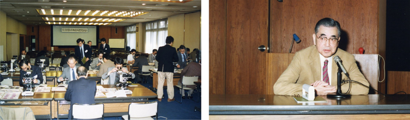
Figure 11: On the left is the venue for the clinical neuropathology conference, and on the right is Professor Shiraki giving a special lecture. Conducting research at Aichi Medical University In 1993, I became a professor at the Institute for Medical Science of Aging, Aichi Medical University. This research institute was founded by Profes-sor Hisashi Tauchi, professor emeritus at Nagoya University and an authority on the pathology of aging. Dr. Tauchi is a pathologist who pioneered pathological research in geriatric medicine. Even after he turned 80, Dr. Tauchi continued to publish pathological research on Japanese centenarians as a book, and he also published a book in English (20). I also wrote an article on the neuropathology of centenarians in that book. Dr. Tauchi was the first in the world to propose that the number of cells decreases with age, causing organ atrophy. Even after being appointed as a professor at the Institute for Medical Institute of Aging, Aichi Medical University, I continued to strive to make the Institute one of the leading neuropathological institutes in Japan. On the left in Figure 12 is the building of the Institute, and the right is Professor Tauchi (marked with an asterisk) with my colleagues at the Institute when I was appointed as a professor.
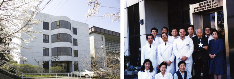
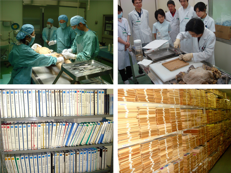
Figure 12: On the left is the building of Institute for Medical Institute of Aging, and on the right is Professor Tauchi (marked with an asterisk) with my colleagues at the time of my appointment. In my third year at the Institute for Medical Science of Aging, Dr. Mari Yoshida, with whom I had previously conducted joint research, joined the Institute. Dr. Yoshida specializes in neurology and has grown to become a leading expert in the neuropathology of neurodegenerative diseases in Japan 21,22,23). Afterwards, she succeeded me as a professor at the Institute for Medical Science of Aging. Figure 13 shows a pathology autopsy in the laboratory on the upper left, cutting of a brain by Dr. Yoshida on the upper right, data on autopsied brains on the lower left, and organized specimens on the lower right.
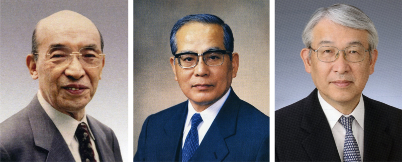
Figure 13: A pathology autopsy in the laboratory on the upper left, cutting of a brain by Dr. Yoshida on the upper right, data on autopsied brains on the lower left, and organized specimens on the lower right. A professor in Nagoya University’s Department of Neurology at that time was Dr. Gen Sobue, who sent many researchers to my laboratory, and we were able to do a lot of joint research. Dr. Sobue is an extremely talented neurologist, active, and he played a major role in elucidating the molecular pathogenesis of ALS and BSMA (24). He is still active as the president of Aichi Medical University. Figure 14 is Professor Itsuro Sobue, Professor Akira Takahashi, and Professor Gen Sobue from the left.
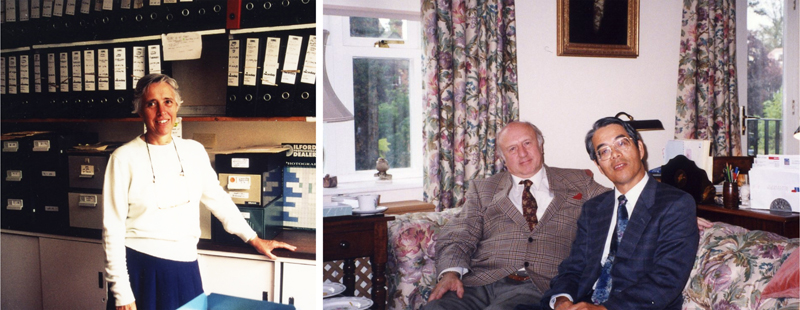
Figure 14: From left, Professor Itsuro Sobue, Professor Akira Takahashi, and Professor Gen Sobue. In 1996, I visited Oxford University, where I was able to study microscopic specimens directly from Professor Margaret Esiri for two months and study at a prestigious neuropathology laboratory. At the time, I was also able to meet Professor J Trevor Hughes, the author of “Pathology of the Spinal Cord” and someone I have long admired. Figure 15 shows Professor Esiri on the left and Professor Hughes and me on the right.
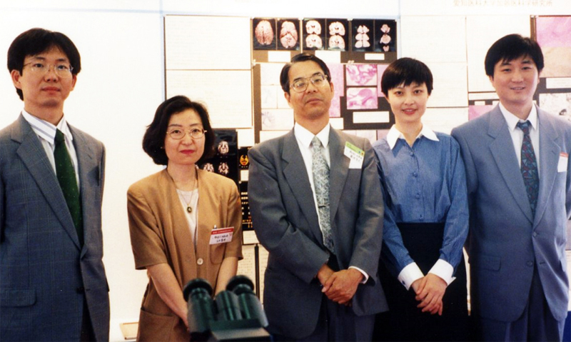
Figure 15: Professor Esiri on the left and Professor J Trevor Hughes and me on the right. I conducted joint research with many young doctors studying neuropathology such as Dr Yasushi Iwasaki, Dr. Maya Mimuro, Dr. Nobuko Ujihira, Dr. Motoko Sakai, Dr. Tamaki Iwase, Dr. Keizo Yasui, Dr. Nozomi Hishikawa, Dr. Yoji Goto, Dr. Junichi Mizuno, Dr. Masumi Ito, Dr. Satoshi Kuru, and Dr. Jun Sone. We also welcomed many researchers from Shanghai. The first person to come to the Institute was Dr. Yin Wang. After coming to Japan, he started studying neuropathology. He worked incredibly hard and made such amazing progress that he obtained his doctorate in medicine in Japan (25). Dr. Wang’s wife is also an excellent physician and has written a wonderful paper. Figure 16 is a photo taken at the meeting of the Japanese Society of Neuropathology. From the left, Dr. Iwasaki, Dr. Yoshida, me, Dr. Yang, and Dr. Wang.
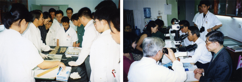
Figure 16: A photo taken at the meeting of the Japanese Society of Neuropathology. From the left, Iwasaki, Yoshida, me, Yang, and Wang. Every year, Dr. Wang referred a neurologist from Shanghai Fudan University. Exchanges with Shanghai continued even after Dr. Wang returned to China. Every year, I visited Shanghai for a research conference, and I was invited as a visiting professor at Fudan University. In 2006, I visited Shanghai, Beijing, and Changchun with many Japanese researchers and became friends with many Chinese professors in each place. I would like to thank Professor Chuanzhen Lu and Professor Zhurong Ye in Shanghai, Professor Luning Wang, Professor Dehong Lu, and Dr. Yueshan Piao in Beijing, and Professor Yu Zhang in Changchun. Dr. Y Wang is currently a professor in Shanghai. The photographs in Figure 17 show a case conference involving group microscopy and cutting of a brain at Fudan University in Shanghai.
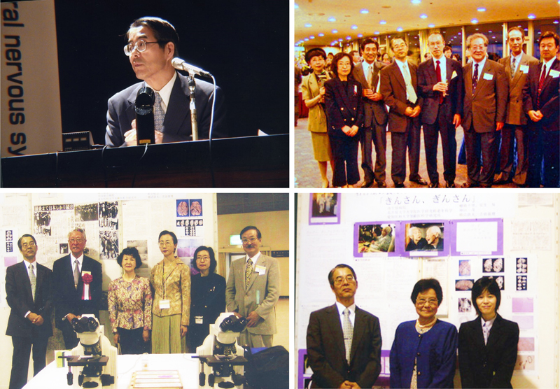
Figure 17: The photograph shows a case conference involving group microscopy and cutting of a brain at Fudan University in Shanghai. In 2003, I hosted the Japanese Society of Neuropathology in Nagoya, where I invited Professor Markus Tolney from Switzerland to give a special lecture on argyrophilic grain disease. At the conference, we were able to hold a symposium on the pathology of spinal cord diseases for the first time as the Japanese Society of Neuropathology. At the conference, we also exhibited the following neuropathological specimens that we had encountered: 1. The brain of Gin-san, a famous twin centenarian from Nagoya and 2. The brain of Shoichi Yokoi, who returned to Japan after living in the jungles of Guam for 28 years after the war and who exhibited Parkinsonism. On the upper left in Figure 18 is me chairing the 44th Annual Meeting of the Japanese Society of Neuropathology. The upper right is the photo at the social gathering of the academic society, the lower left is an exhibit of Mr. Shoichi Yokoi’s pathological specimens, and the lower right is the photo at an exhibit of Gin-san’s pathological specimens.
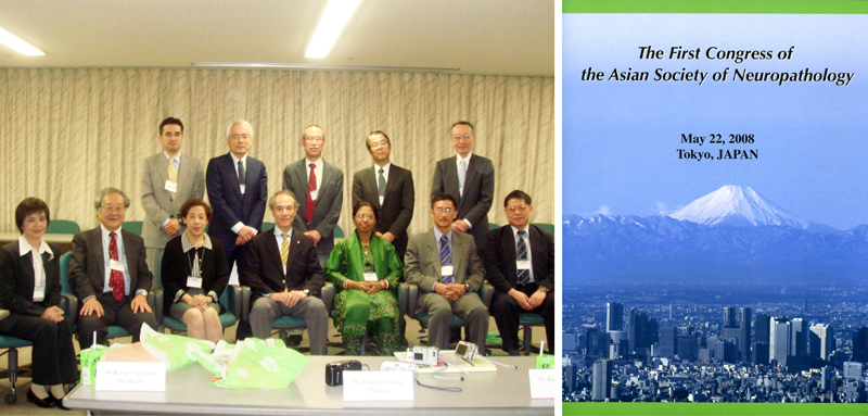
Figure 18: On the upper left is me chairing the 44th Annual Meeting of the Japanese Society of Neuropathology. On the upper right is the photo at the social gathering of the academic society. From the left are doctors Hiroko Yamamoto, Mari Yoshida, Kenji Kosaka, me, Imaharu Nakano, Seung Kim, Kazuo Nagashima, and Mitunori Yamada. On the lower left is an exhibit of Mr. Shoichi Yokoi’s pathological specimens. The third from the left is Mr. Shoichi Yokoi’s wife and the fourth is Dr. Yoko Konagaya, an attending physician of Shoichi Yokoi. On the lower right is a photo at an exhibit of Gin-san’s pathological specimens, the center is Gin-san’s daughter, and on the right is Dr. Chisato Tanahashi, a doctor at Minami Seikyo Hospital who autopsied Gin-san. In addition to researching neuropathology, I also worked to educate medical students at Nagoya University, Nagoya City University, and Aichi Medical University, conducting lectures on neuropathology and providing specimens for pathology training. I continued giving lectures on neuropathology to medical students for about 30 years. In 2007, I happened to be appointed as the 6th president of the Japanese Society of Neuropathology. Based on exchanges with China, I endeavored to hold the first congress of the Asian Society of Neuropathology. The president was Professor Kazuo Nagashima of Hokkaido University, and the vice presidents were Dr. Luning Wang from China and Dr. Seung Kim from South Korea. We succeeded thanks to the cooperation of doctors from various Asian countries. On the left in Figure 19 is a group photo of the organizers of this meeting. The program is on the right.
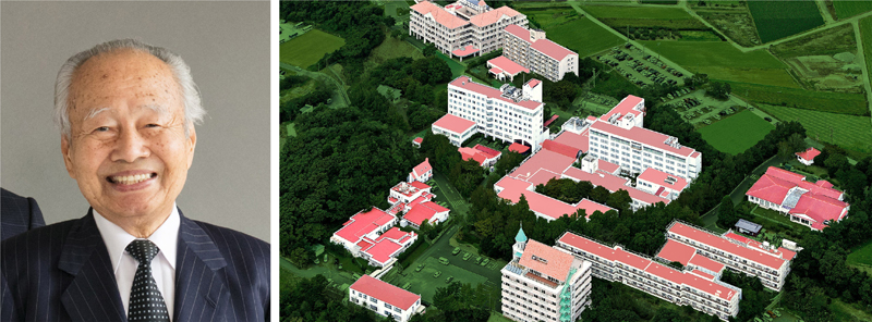
Figure 19: On the left is a group photo of the organizers of the first congress of the Asian Society of Neuropathology. From the left in the front row is Dr. Tumtip Sangruchi from Thailand, Dr. Seung Kim from Korea, Dr. Luning Wang from China, Dr. Kazuo Nagashima, Dr. Chitra Sarkar from India, Dr. Thong Wong from Malasia, and Dr. Chinchen Lee from Taiwan. From the left in the back row is Dr. Masaki Takao, Dr. Atsuo Koto, me, Dr. Imaharu Nakano, and Dr. Yoichi Nakazato. The program of the congress is on the right. We sought to further advance Neuropathology, the bulletin of the Japanese Society of Neuropathology. Its content has been enhanced through the efforts of successive editors-in-chief. As chairman, I helped to enhance a brain bank, which was an effort spearheaded by Dr. Shigeo Murayama of the University of Tokyo. I also strove to establish a regional branch of the Society of Neuropathology. This means that doctors who are interested in neu-ropathology will have the opportunity to study neuropathology wherever they are in Japan. In 2009, the 50th anniversary ceremony of the Japanese Society of Neuropathology was held in Takamatsu under the leadership of President Kiyomitsu Oyanagi. I described the history of the Japanese Society of Neuropathology. The first meeting of the Japanese Society of Neuropathology was held in 1960 in Tokyo, chaired by Professor Tadashi Inose of Yokohama City University. Initially, the principal members of the Society were psychiatrists. Since then, the society has developed, and we were able to hold two international congresses of neuropathology in 1992 and 2018. In 2010, I retired from Aichi Medical University. Afterwards, Professor Yoshida and Professor Iwasaki took over my research. Professor Iwasaki specializes in prion diseases and has produced a number of excellent papers (26). I hope that he will continue to strive to advance the Institute in the future. Thus far, the Institute has assembled over 6,000 autopsied brains with neurological diseases, and our laboratory has grown into one of the leading neuropathological research institutes in Japan. Conducting research at the Institute of Neuropathology at Fukushimura Hospital Since retiring from the Institute of Neuropathology, Aichi Medical University in 2010, I have been working full-time as a doctor at Fukushimura Hospital in Toyohashi. Fukushimura Hospital is a large hospital with 480 beds that specializes in dementia in the Mikawa area. Founded by President Takayuki Yamamoto, the hospital works with many nursing homes and elderly care facilities and is operated by Sawarabikai. On the left side of Figure 20 is President Yamamoto, and on the right side is the full view of Fukushimura Hospital.
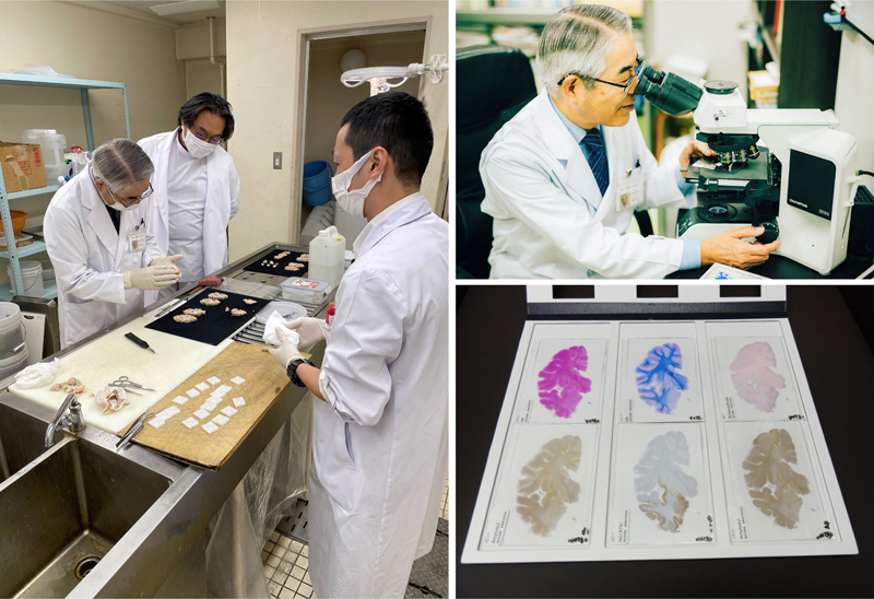
Figure 20: On the left is President Takayuki Yamamoto, and on the right side is the full view of Fukushimura Hospital. President Yamamoto created the Institute of Neuropathology for me in 2010. I am grateful for an environment that allows me to continue working in neuropathology even though I have retired from a university. Fukushimura is a facility that can perform pathology autopsies on nearly 30 dementia patients a year. We hold clinical pathology meetings on three autopsies each month, and we invite many doctors to participate a discussion via Zoom. I am indebted to many people at Fukushimura Hospital, including Director Osamu Kohashi, Vice Director Hiroyuki Ikari, Vice Director Hidechika Okada, Vice Director Yoshiko Yamamoto, Dr. Akira Hori, Dr. Hiroyasu Akatsu, Dr. Keita Sakurai, and technicians Takeshi Kanesaka, Norihiro Ogawa, Naoko Sonoda, and Chieko Taniguchi. The Institute can prepare large specimens of the cerebral hemispheres, allowing us to clearly identify findings that are difficult to gauge in small specimens, such as the spread of lesions and the degree of atrophy. As a facility registered with a brain bank in Japan, we have preserved a large number of frozen tissues from cases confirmed by a neuropathological examination, and we have created a system that allows joint research with researchers here in Japan and abroad. Recently, Dr. Daita Kaneda has joined us as a full-time doctor and he has been very active. On the left in Figure 21 is cutting of a brain at Fukushimura Hospital, on the upper right is me observing a specimen under a microscope, and on the lower right is a large tissue specimen of the cerebral hemisphere.
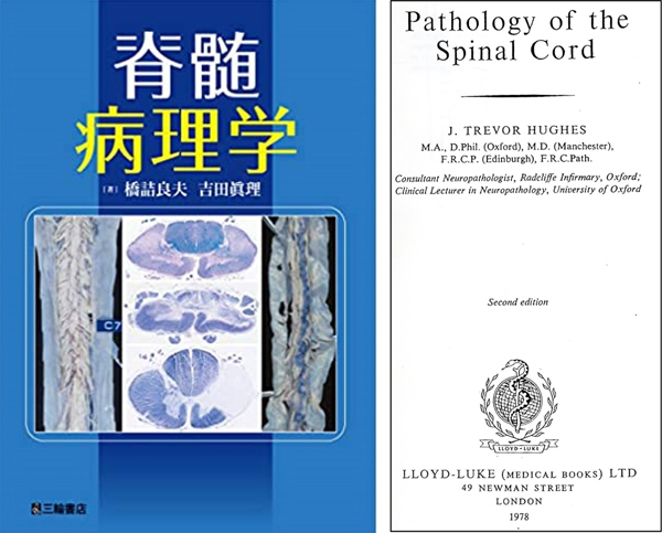
Figure 21: On the left is cutting of a brain at Fukushimuara Hospital. From the left is Dr. Daita Kaneda, me, and technician Takeshi Kanesaka. On the upper right is me observing a specimen under a microscope, and on lower right is a large tissue specimen of the cerebral hemisphere. In 2019, I was able to publish “Spinal Cord Pathology,” co-authored with Professor Yoshida from Miwa Shoten, as the culmination of my research (27). This book describes the pathology findings observed during an autopsy of various spinal cord diseases that occur during the process from human conception to aging. The purpose of this book is to provide useful information for those involved in clinical, pathological, and basic research on spinal cord diseases. This is a collection of 900 valuable histopathology photographs of spinal cord diseases that I have compiled over a long period of time, and I am proud of its unique focus on spinal cord pathology. I was very pleased that Dr. Akira Takahashi, Professor Emeritus of Nagoya University and someone whom I respect, praised my work as “a historical masterpiece.” On the left in Figure 22 is the cover of “Spinal Cord Pathology” and on the right is the cover of “Pathology of the Spinal Cord” by J Trevor Hughes, published in 1978. This is the book that has long been my goal.
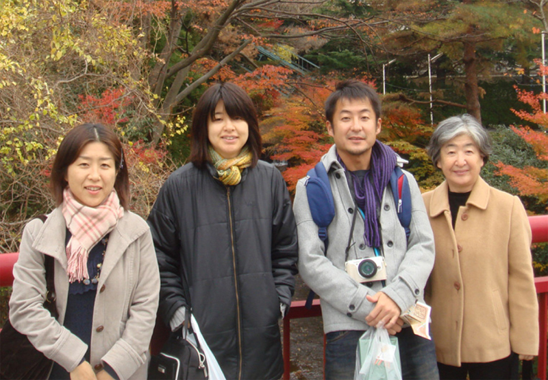
Figure 22: On the left is the cover of “Spinal Cord Pathology,” and on right is the cover of “Pathology of the Spinal Cord” by J Trevor Hughes, published in 1978. This is the book that has long been my goal. In 2018, I was able to give a special lecture entitled the Neuropathology of Neurodegenerative Diseases at the International Congress of Neuropathology held in Tokyo. In this lecture, I was able to present a summary of the neuropathological findings of neurodegenerative diseases that I have been working on for a long time (28). In addition, I was able to make a presentation on observations from a large number of autopsied brains with dementia that I encountered at the Institute of Neuropathology, Fukushimura Hospital at the symposium of the Japanese Society of Neurology in 2019. I was able to publish the content of that presentation as “Macroscopic findings of brain with dementia” in the journal Neuropathology (29). I will be 80 years old in 2024, so I plan to retire from the Institute of Neuropathology at Fukushimura Hospital. Finally, I will list some of the major papers by joint researchers that could not be included here (30-44). In conclusion I am grateful for the good fortune that I have enjoyed to be able to continuously conduct research specialized in neuropathology for 50 years. I am grateful to the many teachers who have supported my research, my colleagues who have conducted joint research with me, the clinicians and pathologists who have been involved in many pathological autopsies, and the technicians who have prepared so many specimens. I would like to end this article by thanking my family. My wife is a volunteer at a cancer palliative care facility, my eldest daughter is a pediatrician, my eldest son is a researcher at a major electronics company, and my second daughter is a physician. I am glad that each of them is actively living a meaningful life. Figure 23 shows my family, from left to right: My eldest daughter Yukiko, my second daughter Mariko, my eldest son Kenichi, and my wife Kiyoko.
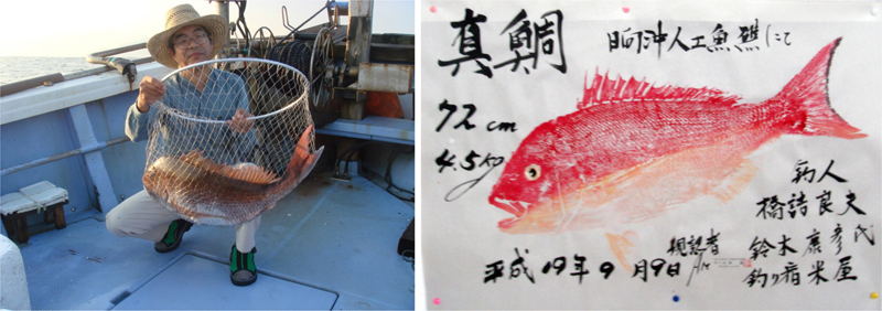
Figure 23: My family, from left to right: My eldest daughter Yukiko, my second daughter Mariko, my eldest son Kenichi, and my wife Kiyoko. As a hobby, I have been boat fishing for more than 40 years, and I go out with my fishing friends to Wakasa Bay and Ise Bay for red sea bream, flounder, and yellowtail. I am happy that I can still drive my own car and attempt to fish. I am grateful for my health. On the left in Figure 24 is me on a fishing boat in Wakasa Bay, and on the right is a print of a red sea bream. Finally, I would like to thank Professor Werner Paulus and Dr. Osamu Yokota, Okayama University for giving me the opportunity to write this article.
Figure 24: On the left is me on a fishing boat in Wakasa Bay, and on the right is a print of a red sea bream. References 1. Hashizume Y. Fünf erwachsene Fälle der von der Ependymitis granularis erregten Aqueductsstenose [5 adult cases of cerebral aqueduct stenosis caused by ependymitis granularis]. Acta Pathologica Japonica. 1976; 27:409-419. PMID: 920176 2. Hashizume Y. Pathological study on the ossification of the posterior longitudinal ligament. Acta Pathologica Japonica. 1980; 30:255-273. https://doi.org/10.1111/j.1440-1827.1980.tb01320.x 3. Hashizume Y, Iijima S, Kishimoto H et al. Pathology of spinal cord lesions caused by ossification of the posterior longitudinal ligament. Acta Neuropathol. 1984; 63:123-130. https://doi.org/10.1007/BF00697194 4. Kameyama T, Hashizume Y, Ando T et al. Spinal cord morphology and pathology in ossification of the posterior longitudinal ligament. Brain 1995; 118:263-278. https://doi.org/10.1093/brain/118.1.263 5. Hashizume Y, Kishimoto H, Iijima S. Occlusion of the basilar artery. A clinical and pathological study of thirteen autopsied cases. Acta Pathologica Japonica. 1984; 34:29-40. PMID: 6730967 6. Hashizume Y, Iijima S, Kishimoto H et al. Pencil-shaped softening of the spinal cord. Pathologic study in 12 autopsy cases. Acta Neuropathol. 1983; 61:219-1224. https://doi.org/10.1007/BF00691989 7. Hashizume Y, Hirano A. Intramedullary spinal cord metastasis. Acta Neuropathol. 1983; 61:214-218. https://doi.org/10.1007/BF00691988 8. Kumagai T, Hashizume Y. Morphological and morphometric studies on the spinal cord lesions in Werdnig-Hoffman. Brain and Development. 1982; 4:87-96. https://doi.org/10.1016/s0387-7604(82)80002-8 9. Riku S, Hashizume Y. A clinico-pathological study on multiple system atrophy, with special reference to its striato-nigral lesions and motor neuron involvements (in Japanese with English abstract). Rinsho Shinkeigaku. 1984; 24:552-561. PMID: 6499329 10. Sakai M, Hashizume Y, Muroga T, Murakami N, Yamamoto H. Neuropathological studies of familial amyotrophic lateral sclerosis with special reference to systemic degeneration of dentato-rubral system and neuronal loss of Onuf’s nuclei (in Japanese with English abstract). Rinsho Shinkeigaku. 1988; 28:1197-1205. PMID: 3219810 11. Hirunagi N, Hashizume Y, Hamaguchi Y, Katsui Y, Kashiwagi H. An autopsy case of Alzheimer’s disease associated with Parkinson’s disease, compared to 2 autopsy cases of diffuse Lewy body disease (in Japanese with English abstract). Nihon Ronen Igakkai Zasshi. 1990; 27:214-219. https://doi.org/10.3143/geriatrics.27.214 12. Okuda S, Ito E, Hashizume Y, Takahashi A. Topographic correlation between basilar artery occlusion and cerebellar involvement--A clinicopathological study (in Japanese with English abstract). Rinsho Shinkeigaku 1991; 31:603-609. PMID: 1934774 13. Kume A, Takahashi A, Hashizume Y, Asai J. A histometrical and comparative study on Purkinje cell loss and olivary nucleus cell loss in multiple system atrophy. J Neurol Sci. 1991 ; 101:178-186. https://doi.org/10.1016/0022-510x(91)90043-7 14. Ando T, Yanagi T, Itoh T, Yamamura A, Takahashi A. Dynamic MR imaging of the cervical cord in patients with cervical spondylosis and ossification of the posterior longitudinal ligament--Significance of dynamic cord compression (in Japanese with English abstract). Rinsho Shinkeigaku. 1992; 32:30-36. PMID: 1628434 15. Inagaki T, Yamamoto T, Hashizume Y et al. Intellectual ability of daily living of centenarians in institutions for the elderly (in Japanese with English abstract). Japanese J Geriat. 1992; 29:849-854. https://doi.org/10.3143/geriatrics.29.849 16. Miyao S et al. Leukoaraiosis in relation to prognosis for patients with lacunar infarction. Stroke. 1992; 23:1434-1438. https://doi.org/10.1161/01.str.23.10.1434 17. Watanabe M, Takahashi A, Hashizume Y, Motegi Y, Furuse M. Magnetic resonance angiography in 12 patients with Wallenberg’s syndrome (in Japanese with English abstract). Rinsho Shinkeigaku. 1992; 32:1186-1192. PMID: 1301318 18. Yoshida M, Murakami N, Hashizume Y, Takahashi A. A clinicopathological study on 13 cases of motor neuron disease with dementia (in Japanese with English abstract). Rinsho Shinkeigaku. 1992; 32:1193-1202. PMID: 1301319 19. Kameyama T, Hashizume Y, Ando T, Takahashi A. Morphometry of the normal cadaveric cervical spinal cord. Spine. 1994; 19:2077-2081. https://doi.org/10.1097/00007632-199409150-00013 20. Tauchi H, Sato T, Watanabe T eds. Japanese Centenarians: Medical Research for the Final Stages of Human Aging. Institute for Medical Science of Aging, Aichi Medical University, 1999. 21. Yoshida M. Multiple system atrophy: Alpha-synuclein and neuronal degeneration. Neuropathology. 2007; 27:484-493. https://doi.org/10.1111/j.1440-1789.2007.00841.x 22. Yoshida M. Cellular tau pathology and immunohistochemical study of tau isoforms in sporadic tauopathies. Neuropathology 2006; 26(5):457-470. https://doi.org/10.1111/j.1440-1789.2006.00743.x 23. Yoshida M. Amyotrophic lateral sclerosis with dementia: The clinicopathological spectrum. Neuropathology. 2004 Mar; 24(1):87-102. https://doi.org/10.1111/j.1440-1789.2003.00544.x 24. Sobue G, Hashizume Y, Mukai E et al. X-linked recessive bulbospinal neuronopathy. A clinicopathological study. Brain. 1989; 112 :209-232. https://doi.org/10.1093/brain/112.1.209 25. Wang Y, Hashizume Y, Yoshida M et al. Pathological changes of the spinal cord in centenarians. Pathol Int. 1999; 49:118–124. https://doi.org/10.1046/j.1440-1827.1999.00832.x 26. Iwasaki Y, Tatsumi S, Mimuro M, Kitamoto T, Hashizume Y, Yoshida M. Relation between clinical findings and progression of cerebral cortical pathology in MM1-type sporadic Creutzfeldt-Jakob disease: Proposed staging of cerebral cortical pathology. J Neurol Sci. 2014; 341:97-104. https://doi.org/10.1016/j.jns.2014.04.011 27. Hashizume Y, Yoshida M. Spinal Cord Pathology. Tokyo, Miwa Shoten. 2019 28. Hashizume Y. Mentors’ Message to Young Neuropathologists: History of clarifying pathogenesis of neurodegenerative disease and development of neuropathology. 19th International Congress of Neuropathology (ICN2018) September 23, 2018 29. Hashizume Y. Macroscopic findings of brain with dementia. Neuropathology. 2022; 42:353-366. https://doi.org/10.1111/neup.12785 30. Kamiya M, Hashizume Y. Pathological studies of aberrant peripheral nerve bundles of spinal cords. Acta Neuropathol. 1989; 79:18-22. https://doi.org/10.1007/BF00308951 31. Ujihira N, Hashizume Y, Takahashi A. A clinico-neuropathological study on brain death. Nagoya J Med Sci. 1993; 56:89-99. PMID: 7898557 32. Hashizume Y, Yoshida M, Wang Y et al. Pathology of spinal vascular disease. Neuropathology 1997; 17:58-66. https://doi.org/10.1111/j.1440-1789.1997.tb00012.x 33. Yasui K, Hashizume Y, Yoshida M et al. Age-related morphologic changes of the central canal of the human spinal cord. Acta Neuropathol. 1999; 97:253-259. https://doi.org/10.1007/s004010050982 34. Hishikawa N, Hashizume Y, Yoshida M, Sobue G. Clinical and neuropathological correlates of Lewy body disease. Acta Neuropathol. 2003; 105:341-350. https://doi.org/10.1007/s00401-002-0651-4 35. Mizuno J, Nakagawa H, Inoue T, Hashizume Y. Clinicopathological study of “snake-eye appearance” in compressive myelopathy of the cervical spinal cord. J Neurosurg. 2003; 99:162-168. https://doi.org/10.3171/spi.2003.99.2.0162 36. Ding ZT, Wang Y, Jiang YP, Hashizume Y, Yoshida M, Mimuro M, Inagaki T, Iwase T.Characteristics of alpha-synucleinopathy in centenarians. Acta Neuropathol. 2006; 111:450-458. https://doi.org/10.1007/s00401-005-0015-y 37. Kizawa M, Mori N, Hashizume Y et al. Pathological examination of spinal lesions in meningeal carcinomatosis. Neuropathology. 2008; 28:295-302. https://doi.org/10.1111/j.1440-1789.2007.00879.x 38. Liu Y, Mimuro M, Yoshida M, Hashizume Y, Niwa H, Miyao S, Ujihira N, Akatsu H. Inclusion-positive cell types in adult-onset intranuclear inclusion body disease: Implications for clinical diagnosis. Acta Neuropathol. 2008; 116:615-623. https://doi.org/10.1007/s00401-008-0442-7 39. Kuru S, Sakai M, Konagaya M, Yoshida M, Hashizume Y, Saito K. An autopsy case of spinal muscular atrophy type III (Kugelberg-Welander disease). Neuropathology. 2009; 29:63-67. https://doi.org/10.1111/j.1440-1789.2008.00910.x 40. Mimuro M, Yoshida M, Miyao S, Harada T, Ishiguro K, Hashizume Y. Neuronal and glial tau pathology in early frontotemporal lobar degeneration-tau, Pick’s disease subtype. J Neurol Sci. 2010; 15;290:177-182. https://doi.org/10.1016/j.jns.2009.11.002 41. Iwase T, Yoshida M, Hashizume Y, Yazawa I, Takahashi S, Ando T, Ikeda T, Nokura K. Intracranial vascular calcification with extensive white matter changes in an autopsy case of pseudopseudohypoparathyroidism. Neuropathology. 2019; 39:39-46. https://doi.org/10.1111/neup.12518 42. Sakurai K, Kaneda D, Morimoto S, Uchida Y, Inui S, Kimura Y, Kato T, Ito K, Hashizume Y. Clinicoradiological features in progressive supranuclear palsy comorbid with argyrophilic grains. Mov Disord Clin Pract. 2022; 9:484-488. https://doi.org/10.1002/mdc3.13455 43. Sakai M, Kuru S, Konagaya M, Kameyama T, Yoshida M. Hereditary spastic paraplegia (in Japanese with English abstract). Spine and Spinal Cord 2022; 35:781-786. 44. Sone J, Ueno S, Akagi A et al. NOTCH2NLC GGC repeat expansion causes retinal pathology with intranuclear inclusions throughout the retina and causes visual impairment. Acta Neuropathol Commun. 2023; 11:71. https://doi.org/10.1186/s40478-023-01564-3
Copyright: © 2023 The author(s). This is an open access article distributed under the terms of the Creative Commons Attribution 4.0 International License (https://creativecommons.org/licenses/by/4.0/), which permits unrestricted use, distribution, and reproduction in any medium, provided the original author and source are credited, a link to the Creative Commons license is provided, and any changes are indicated. The Creative Commons Public Domain Dedication waiver (https://creativecommons.org/publicdomain/zero/1.0/) applies to the data made available in this article, unless otherwise stated. |