Prof. Dr. Michael Schäfers
Multimodal molecular imaging of inflammation
Imaging Technology
Inflammation
Molecular Imaging
Inflammation is involved in many different diseases where it is initiated as a result from various noxes or in consquence of an infection. We develop and apply innovative imaging methodology to visualize the dynamics of molecular players and inflammatory cells (macrophages, neutrophils, T cells) in vivo during the course of disease. Our work involves the identification of inflammatory molecular and cellular targets in inflammation, e.g. matrix-metalloproteinases or S100A8/A9, which can be used for future molecular imaging in vivo. Ligands binding to these targets with both high affinity and specificity are developed, modified and tested in binding assays and cell culture. Ligands are synthetized in-house by a large chemistry and labeling chemistry group and labelled by either radioactive isotopes, fluorescent dyes or photoacoustic absorbers. Our highly interdisciplinary and inter-faculty group at the European Institute for Molecular Imaging is equipped with high-resolution small animal PET/CT, SPECT/CT, optical imaging, photoacoustic and ultrasound systems, small animal surgery, molecular biology labs etc.
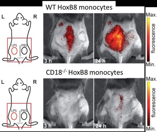
The PhD student project aims at establishing imaging (high-resolution to whole-body) of inflammation in classical immunology models (biopellets, contact dermatitis), autoimmune disease (arthritis) and ischemia-reperfusion (myocardial infarction) to decipher molecular and cellular mechanisms of immune cell responses and their impact on disease course and outcome. The project will employ HoxB8 monocytes in combination with genetic modifications (CRISPR/Cas9) in cooperation with the Institute of Immunology which will be tracked in vivo. The PhD student will be trained in and work with a uniquely broad setup of radioactive and optical state-of-the-art imaging approaches and disease models of inflammation and will combine these with immunohistochemistry, molecular biology and cellular analysis.
Vita
- 1987-1994: Study of Medicine, University of Münster
- 1994-1995: PostDoc, MRC Cyclotron Unit, Imperial College School of Medicine, London, UK
- 1995-1999: Postdoc, Department of Nuclear Medicine, University Hospital Münster
- 2008: Full Professor of Technology and Imaging (W3) and Head of European Institute for Molecular Imaging, University of Münster
- 2011: Coordinator of DFG CRC 656 ‘Cardiovascular Molecular Imaging’
- 2012: Co-Coordinator (together with L. Sorokin and V. Gerke) of DFG Cluster of Excellence EXC 1003 ‘Cells in Motion – CiM’, University of Münster, Germany
Selected references
Gran S, Honold L, Fehler O, Zenker S, Eligehausen S, Kuhlmann MT, Geven E, den Bosch M van, van Lent P, Spiekermann C, Hermann S, Vogl T, Schäfers M and Roth J (2018). Imaging, myeloid precursor immortalization, and genome editing for defining mechanisms of leukocyte recruitment in vivo. Theranostics 8: 2407-2423.
Seifert R, Kuhlmann MT, Eligehausen S, Kiefer F, Hermann S and Schäfers M (2018). Molecular imaging of MMP activity discriminates unstable from stable plaque phenotypes in shear-stress induced murine atherosclerosis. PLoS One 13: e0204305.
Hugenberg V, Wagner S, Kopka K, Schäfers M, Schuit RC, Windhorst AD and Hermann S (2017). Radiolabeled Selective Matrix Metalloproteinase 13 (MMP-13) Inhibitors: (Radio)Syntheses and in Vitro and First in Vivo Evaluation. J Med Chem 60: 307-321.
Gerwien H*, Hermann S*, Zhang X, Korpos E, Song J, Kopka K, Faust A, Wenning C, Gross CC, Honold L, Melzer N, Opdenakker G, Wiendl H, Schäfers M* and Sorokin L* (2016). Imaging matrix metalloproteinase activity in multiple sclerosis as a specific marker of leukocyte penetration of the blood-brain barrier. Sci Transl Med 8(364): 364ra152.
Hermann S, Kuhlmann MT, Starsichova A, Eligehausen S, Schafers K, Stypmann J, Tiemann K, Levkau B and Schäfers M (2016). Imaging Reveals the Connection Between Spontaneous Coronary Plaque Ruptures, Atherothrombosis, and Myocardial Infarctions in HypoE/SRBI-/- Mice. J Nucl Med 57: 1420-1427.
Links