Prof. Dr. Stefan Luschnig
Epithelial tissue dynamics and function
Development
Cell Biology / Molecular Biology
Membrane Protein Dynamics
Tissue dynamics and morphogenesis
Developmental Cell Biology
Advanced microscopy and image analysis
Drosophila genetics and genomics
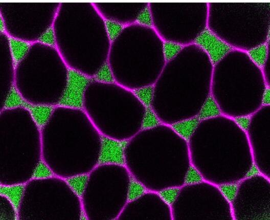
My lab studies the cellular mechanisms that shape developing epithelial tissues and enable physiological functions such as gas exchange, barrier formation, and paracellular transport. We use Drosophila as an accessible model to investigate epithelial tissue dynamics, focusing on the structure and assembly of tricellular junctions (TCJs) and how regulation of adhesion and contractility at vertices controls barrier function. Second, we study how epithelial cells generate tubes of defined size and shape, how sprouting branches invade surrounding tissues, and how epithelial tubes fuse to form interconnected tubular networks during organogenesis. We combine genetics, advanced live imaging, quantitative image analysis, and acute manipulations at the single-cell level in vivo. We have developed genetic tools in Drosophila to manipulate and visualize intracellular trafficking and protein secretion, enabling us to trace secretory routes of cargo proteins and to probe the kinetics of endocytosis and exocytosis in intact tissues. We are recruiting motivated PhD students interested in quantitative developmental cell biology and in vivo imaging to join our collaborative, interdisciplinary team.
Possible PhD projects include
- Dynamics of tricellular junction assembly and remodeling in the follicle epithelium
- Cell biology of tubular morphogenesis
- Analysis of polarized membrane trafficking in epithelial tubes.
Vita
- 1990 - 1996: Studies in Biology, University of Erlangen, Germany
- 2000: Graduation (Dr. rer. nat.) Max-Planck Institute for Developmental Biology, Tübingen, Germany
- 2000 - 2004: Postdoctoral researcher, Stanford University School of Medicine and Howard Hughes Medical Institute, USA
- 2004 - 2007: Research Group Leader, University of Bayreuth, Germany
- 2007 - 2015: Research Group Leader, University of Zurich, Switzerland
- Since 2015: Professor, University of Münster, Germany
Selected references
Jacobs, T., Isasti Sanchez, J., Reger, S., and Luschnig, S. (2025). Rho/Rok-dependent regulation of actomyosin contractility at tricellular junctions restricts epithelial permeability in Drosophila. Current Biology 35(6): 1181-1196. doi.org/10.1016/j.cub.2025.01.043.
Schleutker, R., and Luschnig, S. (2024). Palmitoylation of proteolipid protein M6 promotes tricellular junction assembly in epithelia of Drosophila. Journal of Cell Science, 137(6): jcs261916. doi.org/10.1242/jcs.261916.
Glashauser, J.*, Camelo, C.*, Hollmann, M., Backer, W., Jacobs, T., Isasti Sanchez, J., Schleutker, R., Förster, D., Berns, N., Riechmann, V., and Luschnig, S. (2023). Acute manipulation and real-time visualization of membrane trafficking and exocytosis in Drosophila. Developmental Cell 58(8): 709-723.e7. doi.org/10.1016/j.devcel.2023.03.006.
Camelo, C., Körte, A., Jacobs, T., and Luschnig, S. (2022). Tracheal tube fusion in Drosophila involves release of extracellular vesicles from multivesicular bodies. Journal of Cell Science 135(3): jcs259590. doi.org/10.1242/jcs.259590.
Isasti-Sanchez, J., Münz-Zeise, F., Lancino, M., and Luschnig, S. (2021). Transient opening of tricellular vertices controls paracellular transport through the follicle epithelium during Drosophila oogenesis. Developmental Cell 56, 1083-1099.e5. doi.org/10.1016/j.devcel.2021.03.021.