


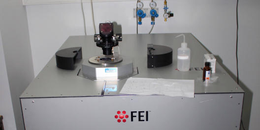
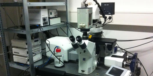
© AG Wedlich-Söldner FEI_1
Location: Institute of Cell Dynamics and Imaging
Group: AG Wedlich-Söldner
Contact Person: Christian Schuberth
Tel.: 0251 83-59053, cschuber@uni-muenster.de
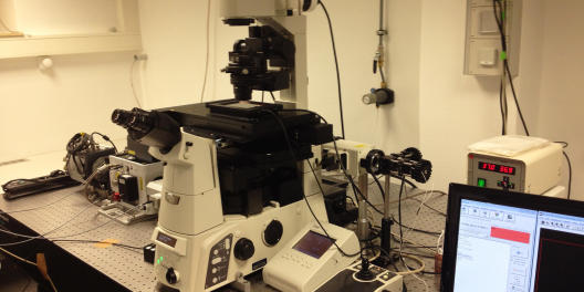
Applications & Info:- Epifluorescence, TIRF, circular TIRF, FRAP
- rapid switch between modes (in ms via Galvos), hardware auto-focus, 405/491/561 nm lasers, automated c-y-z, complex protocols, climate chamber, EMCCD with up to 30 f/s
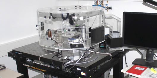
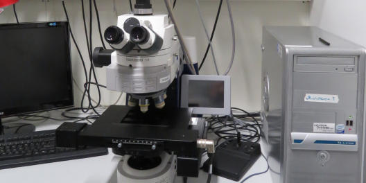
© AG Wedlich-Söldner FEI_2
Location: Institute of Cell Dynamics and Imaging
Group: AG Wedlich-Söldner
Contact Person: Christian Schuberth
Tel.: 0251 83-59053, cschuber@uni-muenster.de
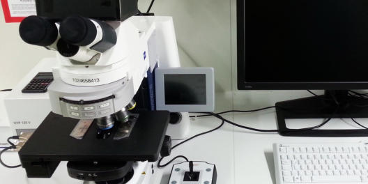
Applications & Info:- Epifluorescence, TIRF, circular TIRF, FRAP, laser ablation
- rapid switch between modes (in ms via Galvos), hardware auto-focus, 405/488/pulsed 355 nm lasers, automated x-y-z, complex protocols, climate chamber, CCD camera
© AG Wedlich-Söldner FEI_3
Location: Institute of Cell Dynamics and Imaging
Group: AG Wedlich-Söldner
Contact Person: Christian Schuberth
Tel.: 0251 83-59053, cschuber@uni-muenster.de
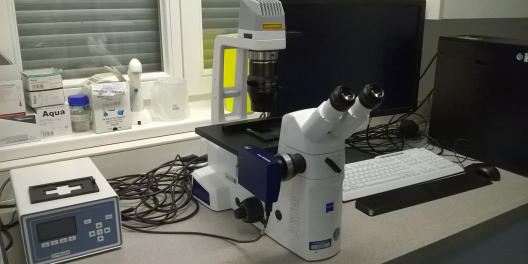
Applications & Info:- Epifluorescence, TIRF, circular TIRF, spinning disk, FRAP
- rapid switch between modes, hardware auto-focus, 405/488/561/640 nm lasers, automated x-y-z, complex protocols, climate chamber, EMCCD with up to 30 f/s (512x512 px, 2x magnification)
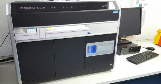
ImageStreamX mkII
Location: Institute of Immunology, Röntgenstraße 21
Group: AG Roth
Contact Person: Achmet Imam Chasa
Tel.: 0251 83-52942, aimam@uni-muenster.deApplications & Info:
- The ImageStreamX is a functional combination of a flow cytometer and a fluorescence microscope.
- It unites the possibility of high throughput data acquisition of flow cytometry with morphological information obtained by microscopy.
- Therefore it is possible to even analyse rare subpopulations of cells for certain expression patterns of target proteins and perform statistical data analyses.
- Examples of possible assays: Transcription factor translocation into nucleus upon cell stimulation; Phagocytosis/endocytosis assay with analysis of subcellular localisation; Protein-protein interaction/co-localisation studies; Cell-cell binding/interactions (APC/T-Cell)
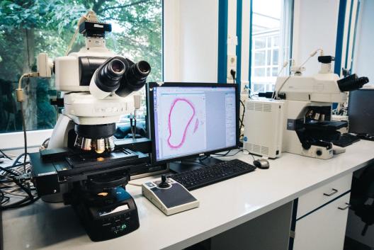
© EIMI Nikon Eclipse Ni-E & OptiGrid structured light illumination system
Location: European Institute for Molecular Imaging, Waldeyerstr. 15
Contact Person: Michael Kuhlmann, Tel.: 0251 83- 49312, kuhlmam@uni-muenster.deApplications & Info:
- Nikon Eclipse Ni-E Microscope for brightfield and fluorescent microscopy
- Motorized desk
- Fully controllable by Nikon NIS Elements AR software package
- 2 camera heads (B/W & RGB)
- Fluorescent filter blocks for DAPI, TRITC, FITC, Cy 5 and Cy 5.5
- automatic scanning of slides in up to 6 dimensions (X,Y,Z, Lambda (wavelength), T, multipoint) possible
- The (optional) OptiGrid converts the illumination system of a conventional widefield microscope to a structured light illumination system and allows for the computer-assisted generation of images of nearly confocal quality.
Nikon Eclipse Ti with Yokogawa X1 Spinning Disc
Location: Institute of Medical Physics and Biophysics, Robert-Koch-Str. 31
Group: AG Galic
Contact Person: Milos Galic
Tel.: 0251 83-51040, galic@uni-muenster.deApplications & Info:
- Confocal Spinning Disc, FRAP/PA unit
- EMCCD camera
Zeiss Axio Imager M2
Location: Institute of Neuropathology, Pottkamp 2
Contact Person: Volker Senner
Tel.: 0251 83-56974, senner@uni-muenster.deApplications & Info:
- “Stereo Investigator” and “Neurolucida” software (MBF bioscience)
- Brightfield, fluorescence (DAPI, GFP, Cy3) and AxioCam MRc - camera
- 2,5x / 5x / 10x / 20x / 40x objectives and motorized stage
Zeiss Vert A1 Fluorescence Cell Culture Microscope
Location: Department of Medicine A, UKM, Building D3, Room 130.082b
Group: AG Berdel
Contact Person: Sebastian Bäumer
Tel.: 0251 83-44811, baumers@uni-muenster.deApplications & Info:
- Inverted cell culture microscope with red/green/Dapi filters and camera soft and hardware
- Heated table

Zeiss Axiovert 200M
Location: Institute of Physiological Chemistry and Pathobiochemistry, Waldeyerstraße 15
Group: AG Sorokin
Contact Person: Sophie Loismann, Tel.: +49 251 83-55583, loismann@uni-muenster.deApplications & Info:
- Inverse light and fluorescence microscope with incubation system and Ibidi pump system connected
- long term live cell imaging for e.g. cell migration and adhesion assays, wound healing/scratch assays (heating/CO2 incubation system attached)
- In combination with pump system: cell alignment under flow, shear stress/flow experiments (Ibidi pump system for long time flow and syringe pump for short time flow/shear experiments)
- Bright field, phase contrast and fluorescence imaging combined with time lapse and/or options for motorized stage (defining several positions per time point or area scan possible)
- LSM800 with Airyscan
Zeiss LSM 900
Location: Institute of Physiological Chemistry and Pathobiochemistry, Waldeyerstraße 15
Group: AG Sorokin
Contact Person: Melanie Hannocks, Tel +49 251 83-55585, hannock@uni-muenster.deApplications & Info:
- Classical confocal microscopy
- Uses the same Zen software and the 3D datasets can be rendered with Volocity
- Excitation lines 405nm, 488nm, 555nm and 633nm
- Objectives: 10x/0.3, 20X/0.8, 40X/1.3DIC, 63X/1.4 Oil DIC
Zeiss LSM 700
Location: Institut für Molekulare Zellbiologie, Schlossplatz 5
Contact Person: Joanna Chiang
Tel.: 0251 83-21761, jchia_01@uni-muenster.deApplications & Info:
- Inverse fluorescence and confocal microscope with incubation system for life cell imaging
- Equipped with Duolink (simultaneous dual channel imaging) and Hamamatsu EM-CCD ImagEM
- Short and long-term live cell imaging for primary neurons and slice cultures (heating/CO2 incubation system)

Zeiss LSM510
Location: Institute of Infectiology (ZMBE), Von-Esmarch-Str. 56
Contact Person: Dr. Christian Rüter
Tel.: 0251 83-56477, rueterc@uni-muenster.deApplications & Info:
- confocal laser scanning microscope LSM510
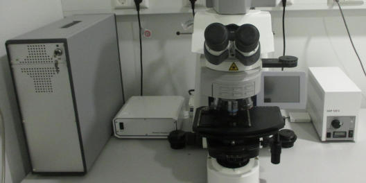
© ZMBE Zeiss Axio imager Z1 equipped with sopt monochrome camera
Location: Institute for Cell Biology
Group: AG Raz
Contact Person: Katsiaryna Tarbashevich
k.tarbashevich@wwu.de, Tel. lab: 0251 83-53018, Tel. office: 83-52183
Applications & Info:- Fluorescence microscopy, FRET maeasurement
- Upright microscope capable of Multi stage imaging, Bright field imaging, Equipped with water-immersive objectives and beam splitter for FRET measurements
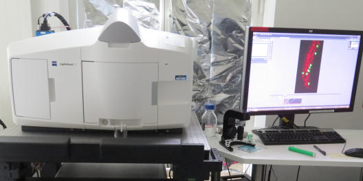
© ZMBE Zeiss Lightsheet Z1 microscope
Location: Institute for cell biology
Group: AG Raz
Contact Person: Łukasz Truszkowski
l_trus01@wwu.de, Tel. lab: 0251 83-58618, Tel. office: 0251 83-52110
Applications & Info:- Fluorescence imaging, lightsheet imaging
- Equipped with water-immersive objectives. The system is suitable for high speed imaging of submerged samples