Welcome to the Near Field Optics group
Group Leader: Dr. Ulrich Fischer

Group Leader: Dr. Ulrich Fischer
Our main research area refers to the use of the optical near field for imaging beyond the diffraction limit.
The most important part of a Scanning Near-Field Optical Microscope (SNOM) is the near field probe which is used as a nanoscopic source of light.
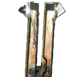
We use glass fragments of a tetrahedral shape with nm- sharp edges and corners as a transparent body of our SNOM probes. To form a SNOM probe (T-tip), the fragment is coated with a thin film of silver, aluminum or gold. Alternatively the fragment is coated with a thick film of aluminum. By squeezing the fragment in a controlled fashion against a glass surface, a triangular aperture is formed in the metal film at the apex of the tip. In this way the Triangular aperture probe (TA probe) is made.
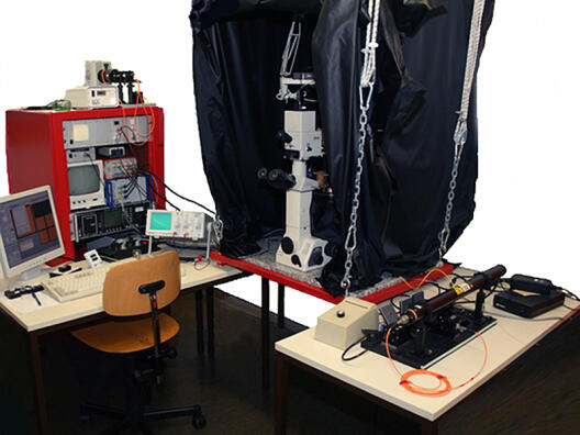
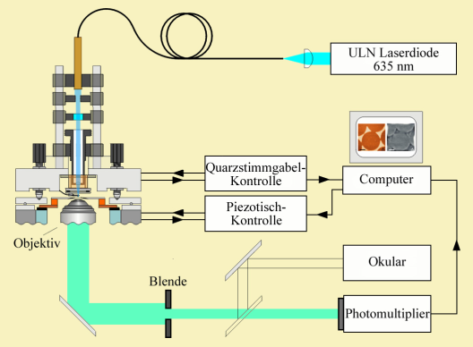
We have developed three different SNOM setups. One microscope uses an auxiliary STM mode for distance control. In a second setup an AFM mode on the basis of quartz tuning forks as a force sensor is used for distance control. Both instruments are operated in a transmission mode. The third setup, also using AFM distance control, can be operated in transmission, internal reflection and PSTM (Photon scanning tunneling microscope) modes.
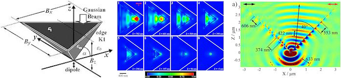
1. The function of the T-tip: Superfocussing of light by a dimensional reduction of surface- to edge- plasmon modes. (K. Tanaka, G. Burr, T. Grosjean, T. Maletzky, U.C. Fischer).
2. Near field microscopy of metal nanostructures: Mapping the surface plasmon mode of metal nanostructures: Mapping the surface plasmon mode of metal nanostructures.
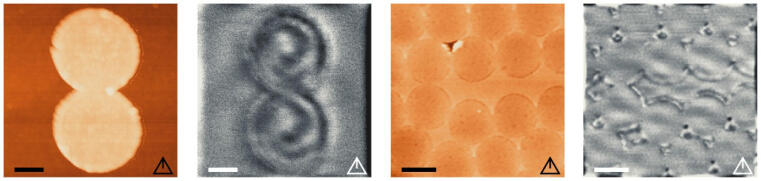
3. Fluorescence Near-Field Microscopy with the T-tip. Previous fluorescence investigations of single molecules were made with the triangular aperture probe showing a resolution of 30 nm. We now started near field fluorescence imaging with the T-tip. We aim at the investigations of Hybrid metal dye nanostructures as artificial antenna complexes and at Near Field Microscopy of Photosynthetic Membrane Systems. (Cooperation with Helmut Kirchhoff, botanical Institute, University of Muenster)
4. Tip enhanced Raman spectroscopy with the T-tip. Development of an imaging mode for mapping the molecular structure of surfaces at a nanoscopic scale.
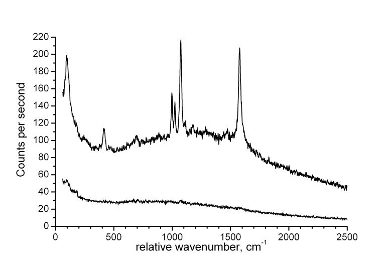
E.G. Bortchagovsky, S. Klein, H. Fuchs, U.C. Fischer. Oral contribution to XXI International Conference on Raman Spectroscopy ICORS 2008 17th - 22nd August 2008 Uxbridge, West London, UK
5. The Fabrication of chemical nanostructures by using nanosphere lithography (NSL).
Originally nanosphere lithography was developed by us to fabricate metal nanostructures as test structures for near field imaging purposes. NSL has become a widely used convenient method for nanofabrication. We extended the use of nanosphere lithography to form chemical nanostructures of multifunctional selfassembled monolayers (NanoMuse).
Cooperation with Stephanie Hoeppener, Laboratory of macromolecular Chemistry and nanoscience, Eindhoven University of technology, Eindhoven, Netherlands
N. Herzer, S. Hoeppener, U.S. Schubert, H. Fuchs, U.C. Fischer 2008 Advanced Materials, 20, 346
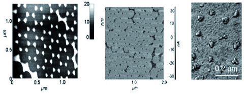
a. Topography of Gold latex bead projection pattern.
b. AFM phase image of a bifunctional nanomuse consisting of a selfassembled monolayer of a silane with a thiol endgroup (triangles)and aliphatic endgroup at other sites.
c. AFM image of gold nanoparticles selectively bound to the thiol groups.
Publication:
Nahfeldoptik - Lichtmikroskopie für die Nanowissenschaften Photonik 5, S.68 (2005)
Cooperations:
Kazuo Tanaka Department of Electronics and Computer Engineering, Gifu University, Gifu city, Japan
Geoffrey W. Burr, IBM Almaden Research Center, San Jose, U.S.A.
Thierry Grosjean, Institut FEMTO-ST, UMR CNRS 6174, Université de Franche-Comté, Besancon, France
Stephanie Hoeppener, Laboratory of macromolecular Chemistry and nanoscience, Eindhoven University of technology, Eindhoven, Netherlands
Alain Dereux, Gérard Colas des Francs, Laboratoire de Physique, Université de Bourgogne, Nanosciences Optique Submicronique Dijon, France
Helmut Kirchhoff, botanical Institute, University of Muenster
Projects:

German Research Foundation (DFG): Near field optics of non radiative surface plasmons at metal nanostructures(terminated)
Tip enhanced Raman spectroscopy and microscopy in the virtual Institute :”Functional properties of aquatic interfaces “ of the Helmholtz Society. (terminated)
Grants of the DFG for E.G. Bortchagovsky and P. Geshev.