Looking Inside the Cell Membrane: How its Properties can be Decoded by Advanced Lasertechnology
Nowadays, lasers are used in many technical contexts. In the medical field, they enable invasive surgery with highest precision. Molecules which are illuminated by laser light do scatter inelastically parts of the light and the shifted frequency of the scattered light can be used as a characteristic fingerprint to identify the type of molecule precisely. This technique is called Raman microscopy and can be exploited as a chemically selective contrast mechanism for marker-free microscopy.
With Raman microscopy, Prof. Carsten Fallnich from the Institute for Applied Physics provides a technical instrument for the study of molecules. In the project presented here, he is working together with Prof. Volker Gerke from the Institute of Medical Biochemistry in order to investigate the structure and behavior of cellular membranes. Earlier analysis methods already indicated that the membrane with its mosaic-like mix of lipids and proteins is highly dynamic and may change depending on the need to take over many functions of the cell. How exactly these processes do take place, and how the building blocks of the cell membrane are arranged in response to certain stimuli is only beginning to emerge. With the high resolution Raman microscopy, Volker Gerke and Carsten Fallnich adress these questions and therby try to advance the field of basic research.
Photos

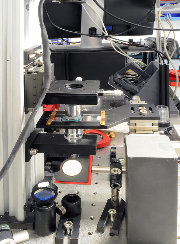
Professor Volker Gerke from the Institute of Medical Biochemistry collides with his microscope to the limit of what can be depicted, because the lipids and proteins that make up the membrane are tiny with their size of six to ten nanometers.© CiM - Elisabeth Deiters-Keul 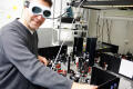
In a joint project with Professor Carsten Fallnich from the Institute of Applied Physics, a technique is used, which can take images even in this nanoscale. The technique is called Raman microscopy.© CiM - Frieda Berg 
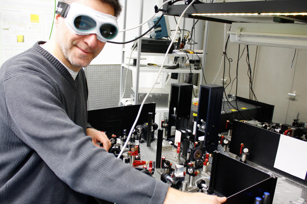
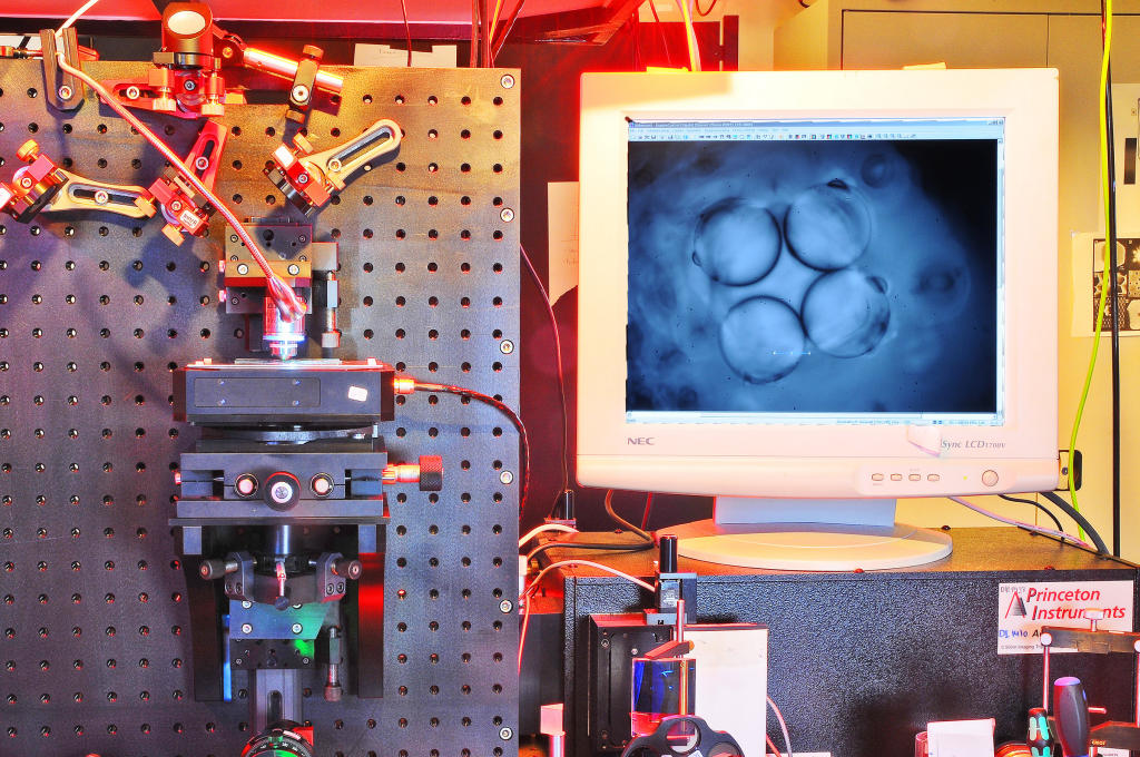
The object is detected by a laser beam which is passed through a complex apparatus.© CiM - Elisabeth Deiters-Keul 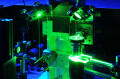
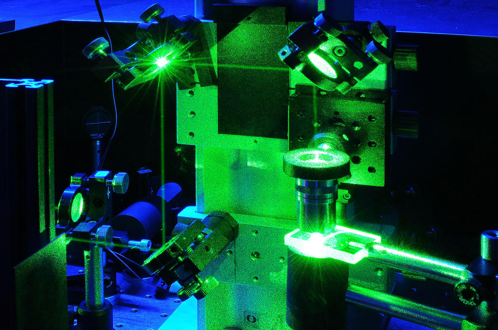
The trick of the Raman microscope: the laser light is scattered by the molecule and sends a specific frequency, quasi its own fingerprint back - every single molecule can be read by this information such as proteins and lipids in the cellular membrane.© CiM - Elisabeth Deiters-Keul 
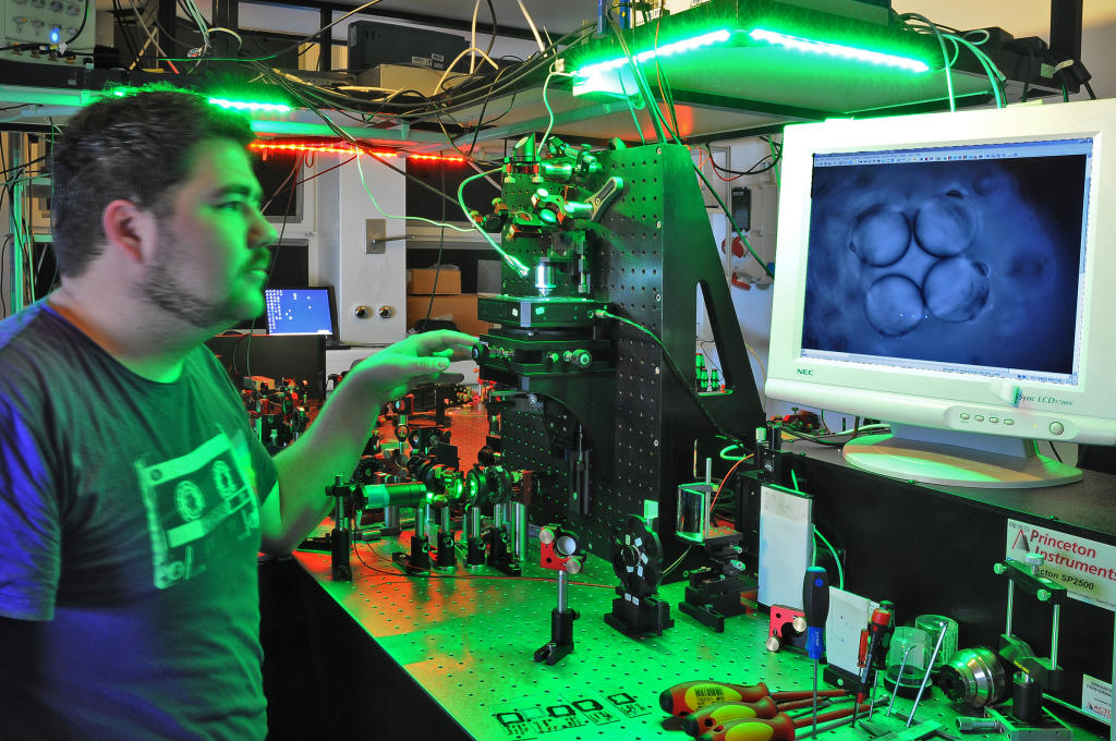
The cooperation of Medical Biochemistry and Applied Physics should make it possible in the future to capture the membrane structure of different cell types in order to see which defects affect membrane function.© CiM - Elisabeth Deiters-Keul