Inflammation in the Brain: Imaging to Investigate Multiple Sclerosis
Published in 2014.
Between the central nervous system that is located in our brain on the one side and a human's blood cirulation on the other, there's a natural border known as the brain-blood barrier. This barrier regulates what gets into the brain and what doesn't.
Multiple sclerosis is an autoimmune disease where an inflammation in the brain develops, while the cause is still not known. Why and how is it possible to have inflammations beyond the brain-blood-barrier? In their project, Prof. Lydia Sorokin and Prof. Günter Haufe want to have a closer look on what's happening inside the brain during multiple sclerosis. How does the disease develop and grow? Therefore, the team applies molecular imaging in order to get significant images without any invasive treatment. With their work, Günter Haufe and Lydia Sorokin contribute to an improved understanding of multiple sclerosis.
Photos

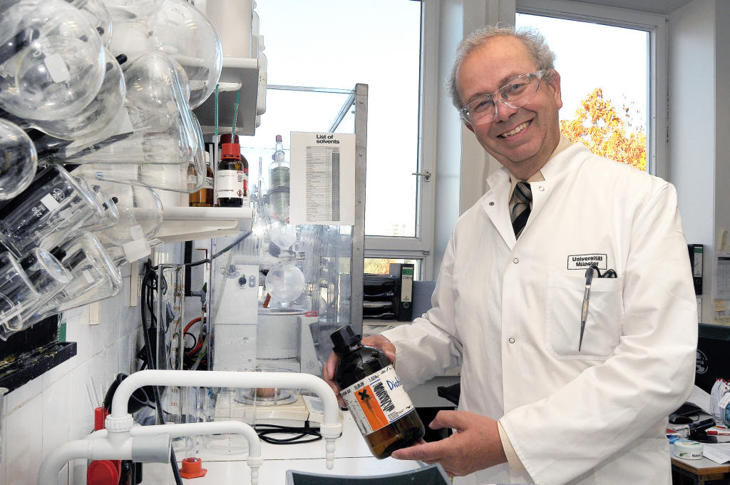
These new images from the brain can be taken with molecular imaging. Therefore, new tracer molecules have to be developed. Professor Günter Haufe is specialist in this field. With his tracers, diseased tissue can be detected under the microscope. Professor Günter Haufe and his team work on the new tracer in the Department of Organic Chemistry. Provided with the knowledge about specific molecular processes happening during a multiple sclerosis attack, the tracer can be designed.© CiM - Elisabeth Deiters-Keul 
PhD student Vysakh Prasad logs the exact composition of the tracer substance. The molecules in the laboratory must fit like a key into the existing lock – the tracer molecules have to bind to such molecules that are involved in multiple sclerosis. © CiM - Elisabeth Deiters-Keul 
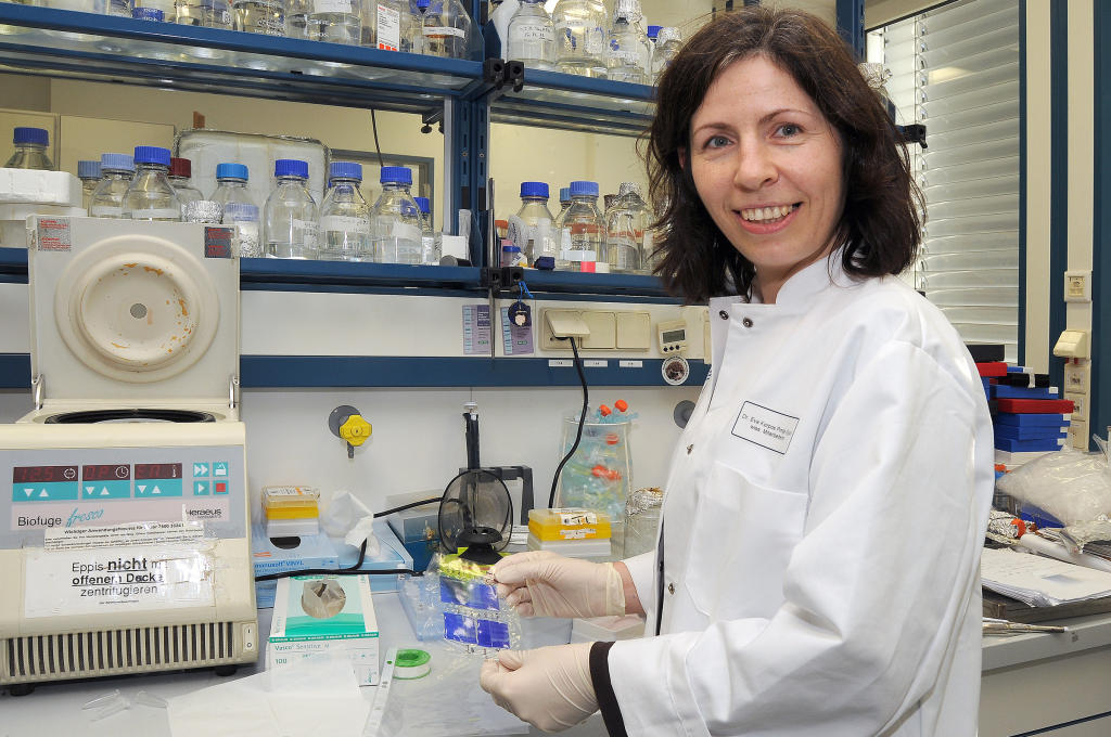
In the next step, first generations of the tracer are applied and tested on tissue sections in the laboratory of Lydia Sorokin, here by Dr. Eva Korpos.© CiM - Elisabeth Deiters-Keul 
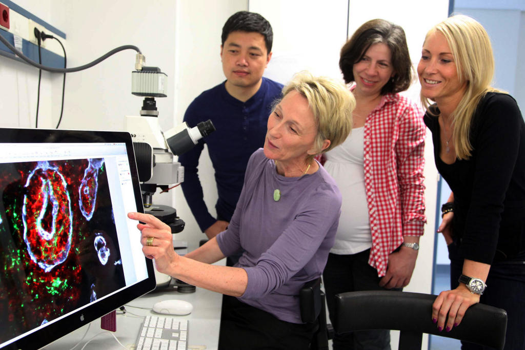
With her colleague Dr. Jiang Song, Eva Korpos evaluates the test results. The aim is to develop the new tracer substance to such an extent that it can be used for the examination of patients in the future.© CiM - Elisabeth Deiters-Keul