
Transmitting stimuli in nerve cells
Nerve cells in the brain communicate by means of special messenger substances, so-called neurotransmitters. They transfer signals from cell to cell. However, this process only occurs when the connections between two cells, the synapses, have enough neurotransmitters at the right places at their disposal. In the case of stimulating nerve cells, this is glutamate – which is otherwise known primarily as an flavour enhancer. The body’s own glutamate, by contrast, is one of the most important messenger substances in the central nervous system. Nerve cells store glutamate in small vesicles in synapses. When nerve cells transmit stimulating signals to other cells, they secrete glutamate in their synapses which electrically stimulates the other receiving cells. Researchers at the Cells-in-Motion Cluster of Excellence at the University of Münster have now discovered how a protein – a so-called glutamate transporter – fills the small vesicles in cell processes with glutamate.
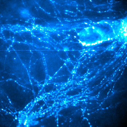
For their study, a team of researchers led by biophysicist Prof. Jürgen Klingauf have, for the first time, observed the process whereby the vesicles are filled with glutamate in living cells. They did so by means of so-called live-cell imaging, using microscopy of those living cells. They were able to show that the amount of glutamate which gets into the vesicles of cell processes depends above all on the chloride content within the vesicles. Researchers had already suspected that there was a connection between chloride and glutamate in the vesicles-filling process in synapses. The Münster researchers are now the first to be able to prove this hypothesis in living cells of mice.
Chloride is present in synapses in the form of sodium chloride – i.e. a salt solution – and transmits electrical signals. This new study shows that when the vesicles are being filled with glutamate, the same amount of chloride flows out into the so-called extra-cellular space. Without this, the absorption of glutamate would not be possible. What is decisive for this exchange process involving glutamate and chloride is a protein, the so-called vesicular glutamate transporter (VGLUT). This glutamate transporter replaces chloride ions with glutamate, and the process is driven by protons with the proton pump H+-ATPase. “Glutamate transporters work like a pump, simultaneously pumping glutamate into the vesicles and removing from them chloride and protons,” explains Jürgen Klingauf. Without the glutamate transporter, no glutamate would get into the vesicles. The results of the study have been published in “Nature Communications”.
Filling the vesicles with glutamate in all stimulating synapses is decisive for how well our nervous system functions. The more the vesicles are filled with glutamate, the bigger the electrical response from synapses can be to a signal, and the better the synaptic connection is between two cells. This in turn has an effect on how good someone’s memory is or how well they can process information. If, however, there is an imbalance in the amount of chloride inside and outside the cell, the glutamate transporter can pump less of the body’s own glutamate into the vesicles in the cell processes. Researchers suspect that there is a connection between this imbalance in the amount of chloride and diseases such as schizophrenia, Alzheimer’s, Parkinson’s and epilepsy. The results of the study are a first indication that this assumption may be true.
The story in detail:
Previous studies had only examined the flow of glutamate in vitro – in other words, not in living nerve cells. It was known that chloride can influence the flow of glutamate by means of the glutamate transporter. It was not, however, possible to prove that the chloride is no longer in the vesicles at the end of this process. The Münster researchers carried out their first experiments in living nerve cells of mice and were thereby able to observe that the membrane of the vesicles is permeable to chloride. “This outflow is decisive for the exchange of chloride and glutamate in the vesicles,” says Jürgen Klingauf.
For their investigations the researchers developed further two so-called fluorescence probes – high-resolution, fluorescent proteins sensitive to chloride and protons. The researchers genetically modified the nerve cells in such a way that the cells produced it themselves. During subsequent experiments with the living nerve cells, the probes are attached to a part of the synaptic vesicles and glowed to different degrees, depending on the number of chloride ions or protons surrounding them. This enabled the researchers to measure how much chloride and protons were contained in a synaptic vesicle.
In developing the fluorescence probes, there were two particular challenges. As the number of chloride ions is so small, it is fundamentally difficult to capture enough light signals. Also, the researchers had to correctly calibrate the actual light signals measured, i.e. correlate the strength of the light to the number of ions measured. In this process, the signals are extremely low.
There was a further challenge before the experiments were started: not every synapse uses glutamate as a messenger substance. Some synapses use the messenger substance GABA. These GABA synapses reduce the stimulation of the nerve cells. “This means we had to separate out these synapses for our investigations,” says Jürgen Klingauf. Using a further fluorescence probe – an antibody against GABA transporters – the researchers were able to find BAGA synapses and eliminate them from their samples.
The Münster researchers had their results confirmed by Prof. Christoph Fahlke from Forschungszentrum Jülich. Fahlke is a specialist for so-called electrophysiological measurements and he and his team were able to measure the electrical chloride flow in the glutamate transporter. Both chloride and glutamate have a negative charge. With their measurements, the Jülich team were also able to demonstrate the flow of charges in both directions, thereby confirming the findings in Münster.
The study received funding from the Cells-in-Motion Cluster of Excellence at the University of Münster, from the Interdisciplinary Centre for Clinical Research (IZKF) at Münster University, from the European Commission and from the German Research Foundation (DFG).
Original publication:
Martineau M, Guzman RE, Fahlke C, Klingauf J. VGLUT1 functions as a glutamate/proton exchanger with chloride channel activity in hippocampal glutamaergic synapses. Nat Commun 2017;8: 2279. Abstract