Online exhibition
Exhibition

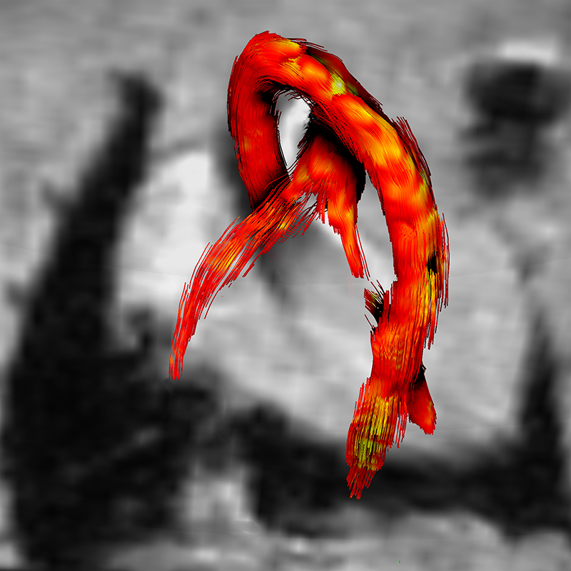
Blood flow in the large arteries. The streamlines display the shape of the blood vessels, and their colour coding (red-orange) depicts the temporal and spatial dynamics of blood flow. 4D phase magnetic resonance imaging© Philipp Bovenkamp, Florian Lindemann, Tobias Brix, Verena Hörr 
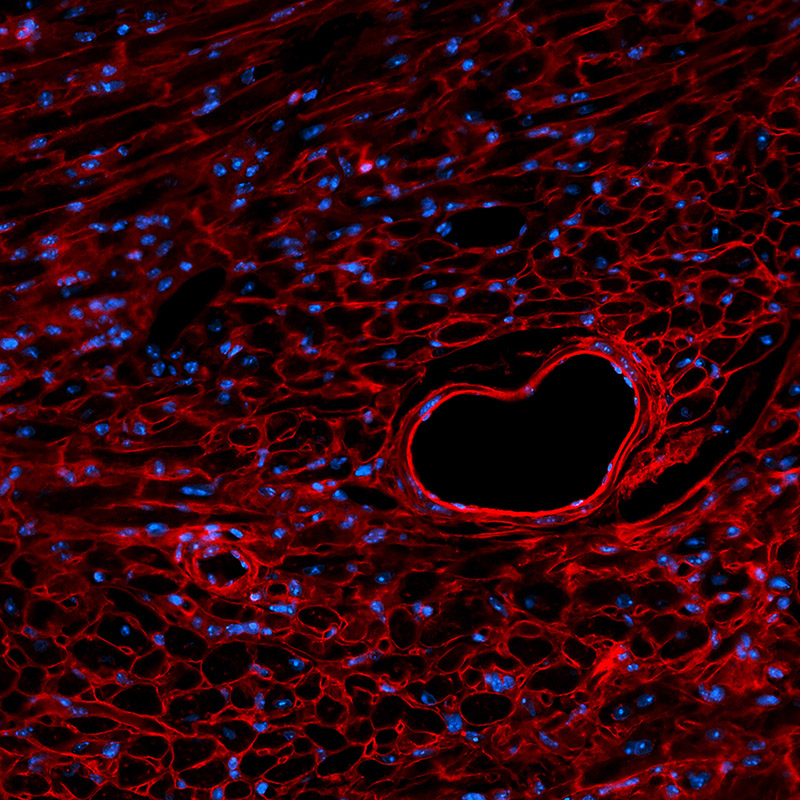
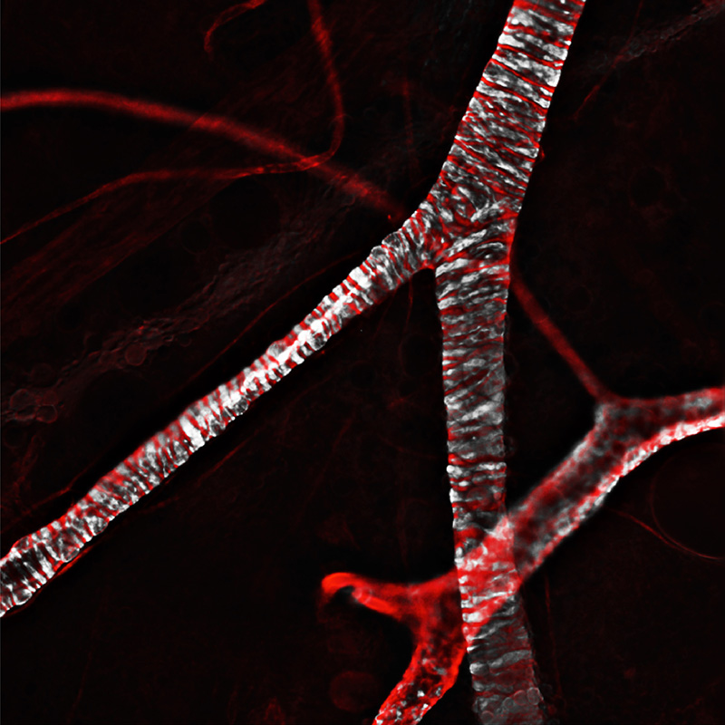
Through arteries and arterioles (foreground) the heart pumps blood throughout the body. It flows back to the heart through venules and veins. The vessels’ muscle cells (white) help to control blood pressure. Confocal fluorescence microscopy© Jacopo Di Russo, Lydia Sorokin 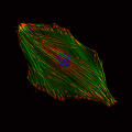
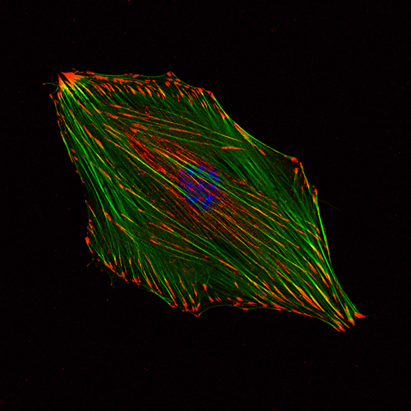
Flexible actin filaments (green) enable a blood vessel’s muscle cells to contract. At the tips of the filaments (red), the cell senses stimuli and reacts by contracting and relaxing synchronously with its neighbouring cells. Fluorescence microscopy© Lema Yousif, Lydia Sorokin 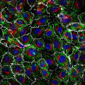
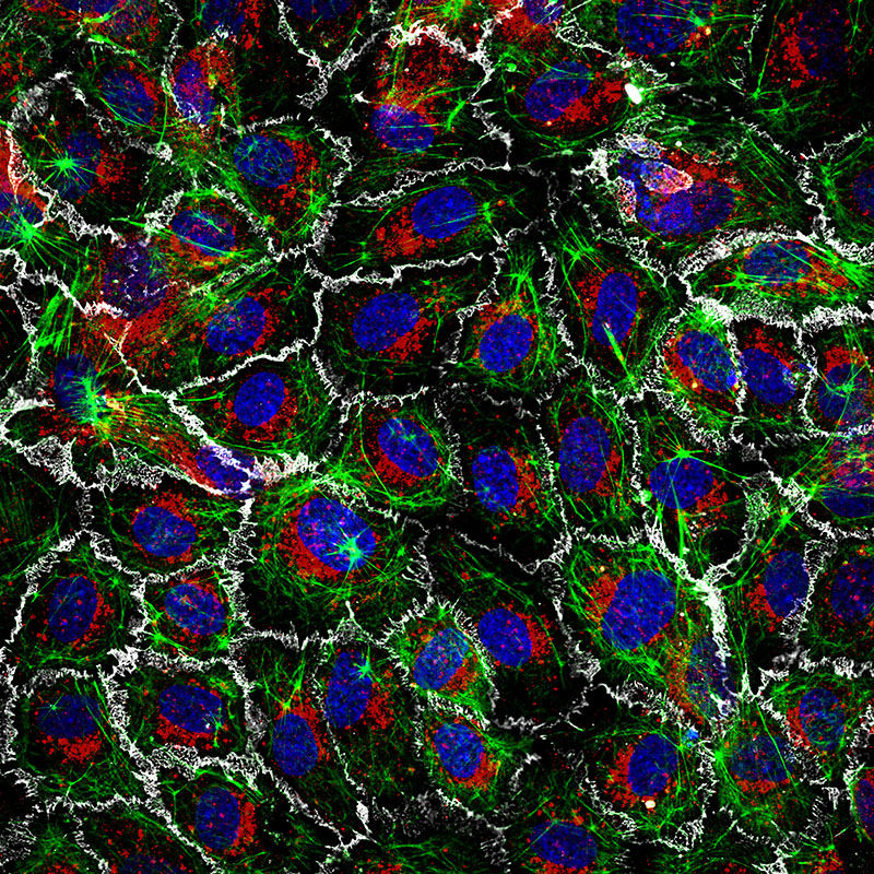
The innermost layer of vessels consists of endothelial cells (blue nuclei, green skeleton). The cells establish specific connections with each other (white), thus regulating material exchange between blood and tissue. Confocal fluorescence microscop© Benjamin Brinkmann, Birgitta Michels, Volker Gerke 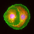
These two blood vessel cells (endothelial cells, pink nuclei) attach tightly to each other. Like glue, the protein integrin (green) connects the membranes of individual cells and transmits signals from one cell to another. Fluorescence microscopy© Sandra Wegner, Rupert Hallmann 
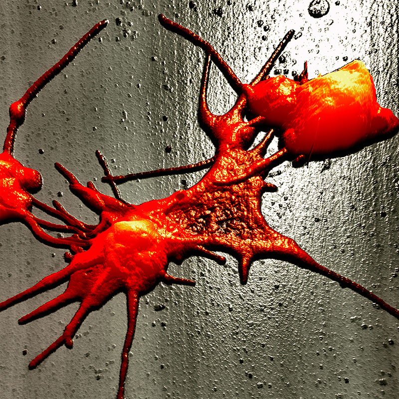
When blood vessels are injured, blood platelets seal the leak. They change their shape, developing protrusions which allow them to stick to the blood vessel wall and establish the first wound closure. Atomic force microscopy© Hermann Schillers, Hans Oberleithner 
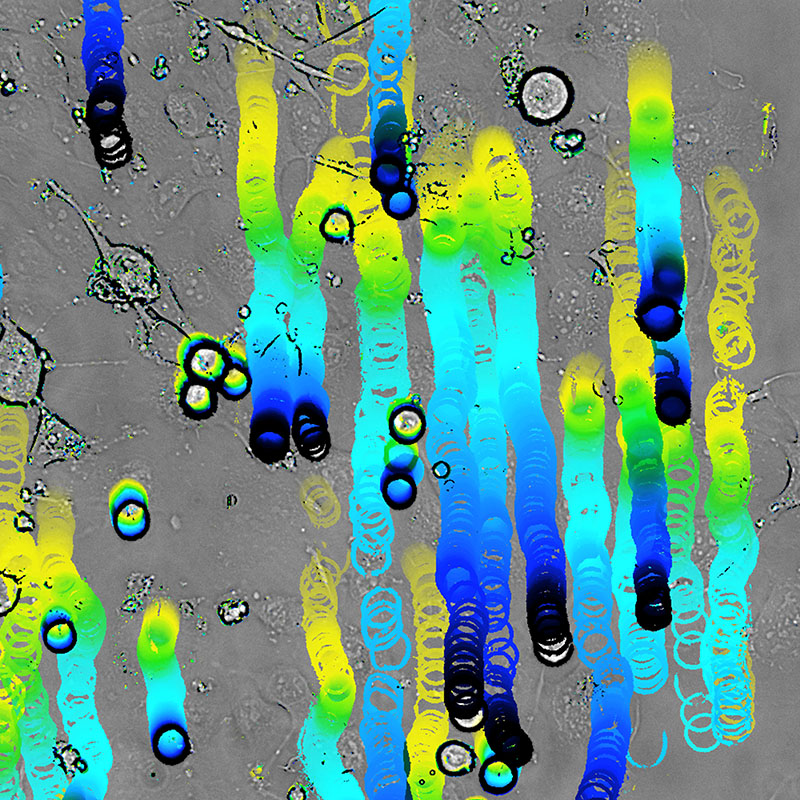
When inflammation occurs, immune cells migrate into the tissue. They roll along the vessel wall (passage of time from yellow to black) and attach to endothelial cells, so that they cannot be washed away by the flowing blood. Bright-field microscopy© Jana Zimmermann, Rupert Hallmann 
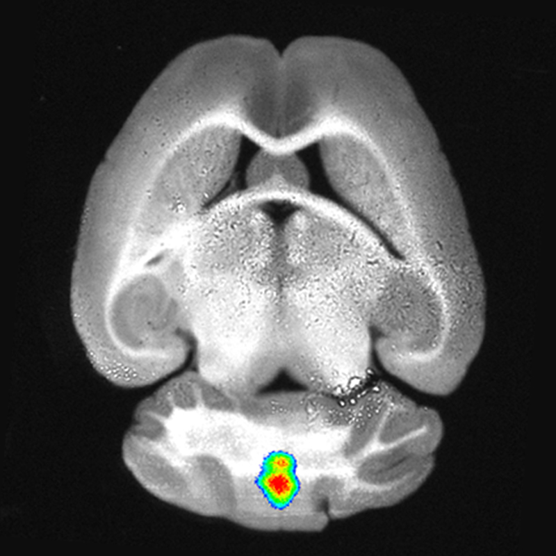
In atherosclerosis, plaque deposits (yellow) build up in arterial walls, which, upon rupture, can lead to heart attack or stroke. CiM scientists investigate inflammatory processes pivotal to this rupture. Planar illumination fluorescence microscopy© Jan Prodöhl, Lisa Honold, Friedemann Kiefer, Michael Schäfers 
Focussed laser light can hold and move small particles, which helps scientists investigate cells. Here, red blood cells (red) in the vascular system of a living zebrafish are trapped in the focus of a laser beam (green). Bright-field microscopy© Robert Meißner, Álvaro Barroso, Christina Alpmann, Cornelia Denz 
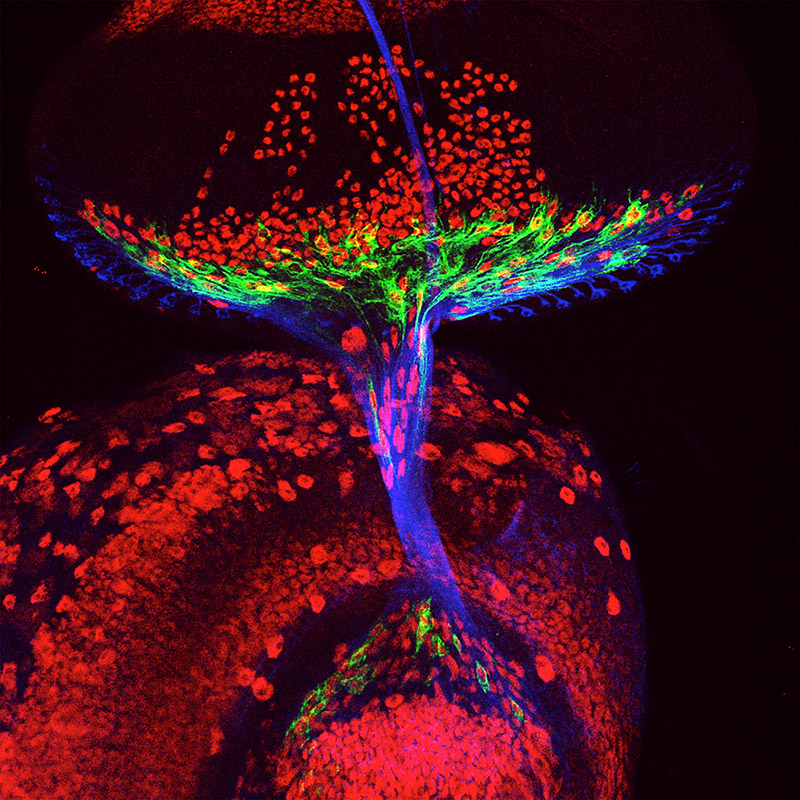
Developing nervous system of a fruit fly. Glial cells (green) migrate along protrusions of nerve cells (blue) from the eye (top) towards the brain (bottom). Both cell types develop in close interaction. Confocal fluorescence microscopy© Anni Bauke, Christian Klämbt 
Sensory nerve cells (green) in the skin of the larva of a fruit fly. Flies undergo a metamorphosis when developing into adults. CiM researchers investigate how cells restructure during this process. Confocal fluorescence microscopy© Svende Herzmann, Sandra Rode, Sebastian Rumpf 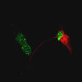
Astrocytes are cells in the brain and spinal cord that contribute to healthy brain activity. The image shows their cell nuclei (green), and a protein (glial fibrillary acidic protein, red) that provides structural support. Confocal fluorescence microscopy© Eva Korpos, Lydia Sorokin 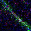
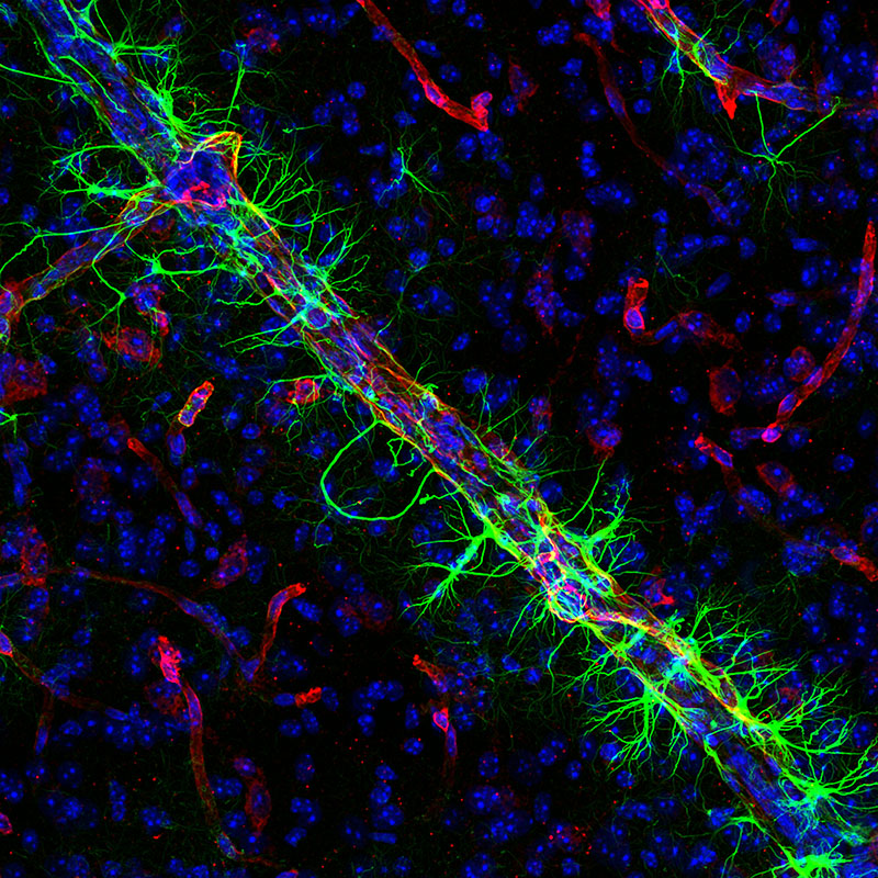
Astrocytes (green) wrap around blood vessels in the brain (red). Their thorny protrusions help form the protective barrier between the nervous tissue and the blood stream: the blood-brain barrier. Confocal fluorescence microscopy© Marcos Assis Nascimento, Lydia Sorokin 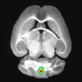
In multiple sclerosis, immune cells manage to migrate into the brain and damage the nerve fibres. Activated enzymes (red, here in a mouse) help the immune cells cross the blood-brain barrier. Fluorescence reflectance imaging© Hanna Gerwien, Andreas Faust, Lydia Sorokin, Michael Schäfers 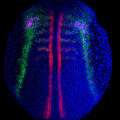
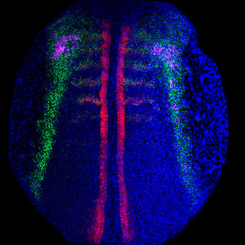
This image shows a twelve-hour-old, yet sexless zebrafish. In the body segments (red), the precursors of germ cells (pink) are forming. A specific attractant (chemokine, green) guides the cells to their destination. Confocal fluorescence microscopy© Azadeh Paksa, Theresa Gross-Thebing, Erez Raz 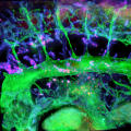
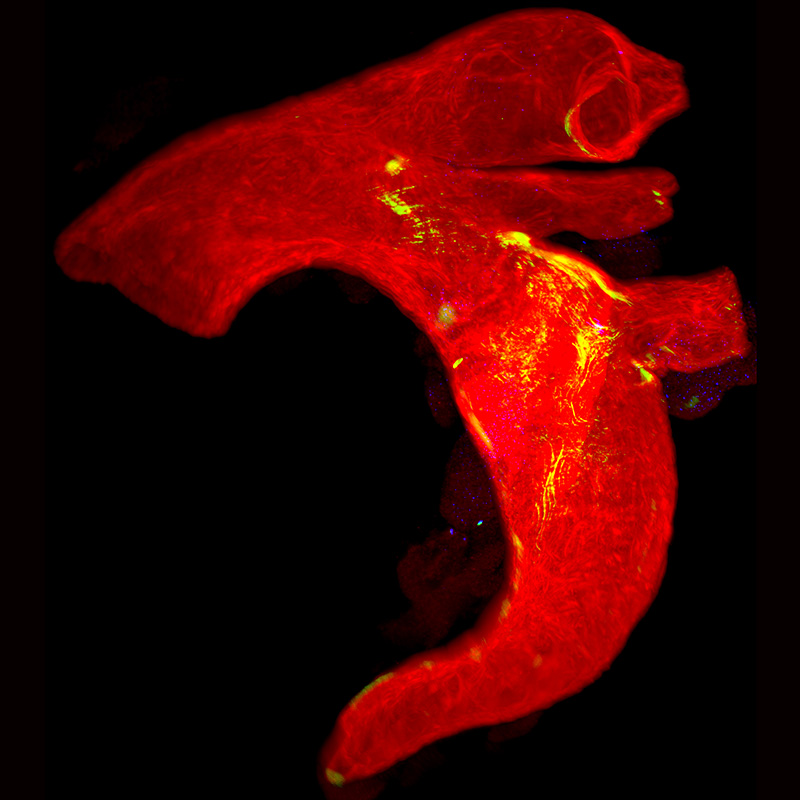
Blood vessels (green), and lymphatic vessels form the transport pathways of the body. In order to form new lymphatic vessels, progenitor cells must migrate away from the veins (blue). Optical sectioning using ultramicroscopy© René Hägerling, Cathrin Pollmann, Friedemann Kiefer 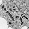
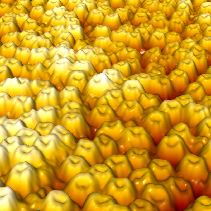
Mitochondria (bean-like structures) are the powerhouses of a cell. Their inner, folded membrane produces substructures called the antechamber (white) and the main chamber (black), thus creating zebra-like stripes in the cross section. Electron microscopy© Stefanie Oeding, Petra Nikolaus, Ulrike Honnert, Martin Bähler 
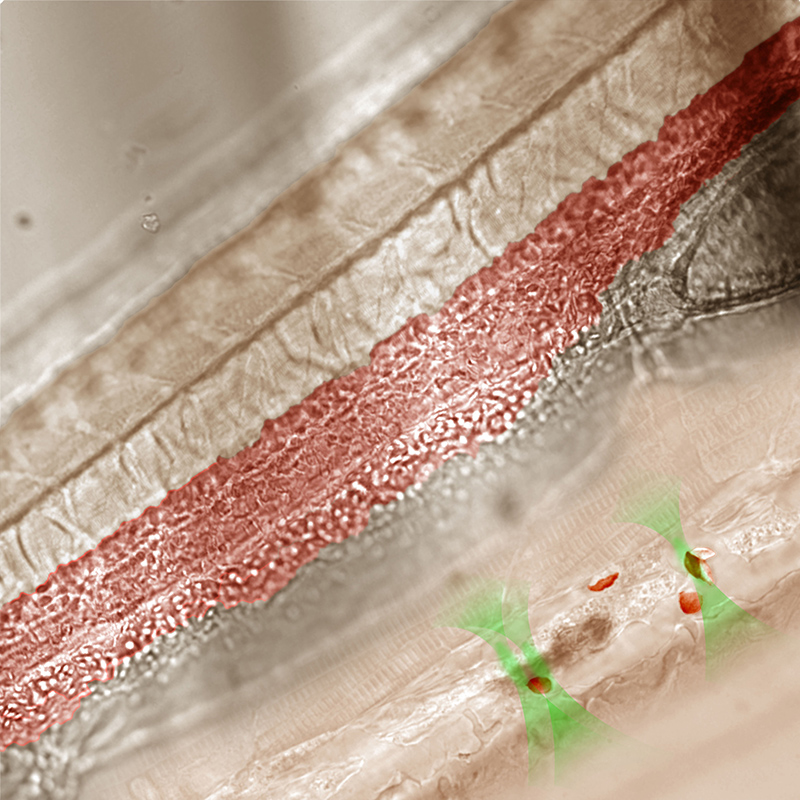
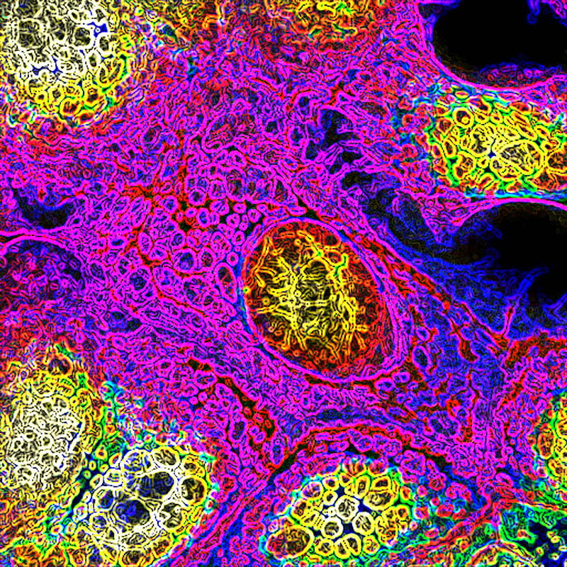
There are millions of nuclear pores on the surface of a claw frog’s egg cell. CiM researchers investigate which factors regulate the width of the pores and thus the admission of proteins. Atomic force microscopy© Victor Shahin, Hans Oberleithner 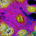
These breast cancer cells are under stress. Calcium is the trigger, and CiM researchers investigate the effects of this stimulus. They want to understand which factors influence the cells’ properties. Confocal fluorescence microscopy© Pauline Wales, Roland Wedlich-Söldner 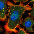
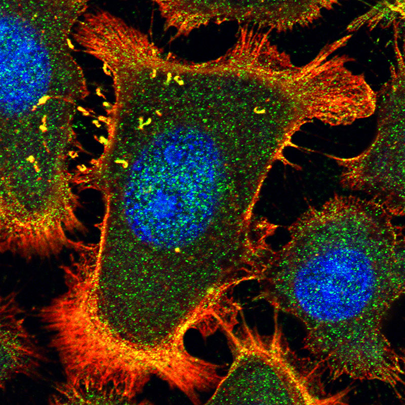
Skin cancer cells move forward by sending out protrusions. In doing so, they continuously rearrange their cytoskeleton (actin, red). Thus, the cells can metastasise from the primary tumour into distant tissues. Confocal fluorescence microscopy© Florian Heßner, Volker Gerke, Ursula Rescher 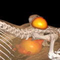
CiM researchers develop methods to visualise cells in the body. Tumour cells (top, yellow-red) avidly take up a radioactive sugar injected into the blood stream. The sugar is excreted through the bladder (bottom). PET-CT© Sven Hermann, Michael Schäfers 
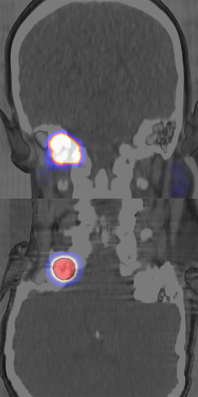
A tumour at the skull base. For cancer diagnosis and therapy, doctors use radioactive substances. These attach themselves to proteins within tumour cells and can be visualized (glowing). Certain types of radiation can destroy tumours (bottom). PET/SPECT-C© Kambiz Rahbar, Lars Stegger, Matthias Weckesser, Michael Claesener, Michael Schäfers 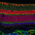
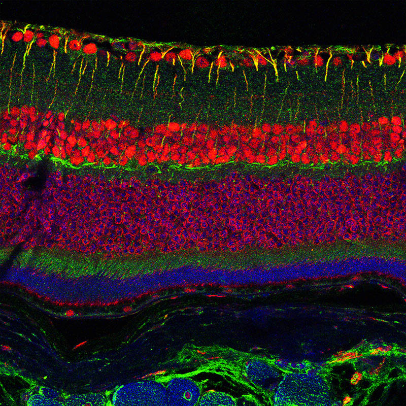
The retina is constructed in layers from the inside (top) to the outside (bottom). CiM researchers investigate whether a receptor of the messenger VEGF (red) is involved in degeneration processes of the retina. Confocal fluorescence microscopy© Anne F. Alex, Nicole Eter 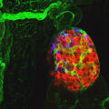
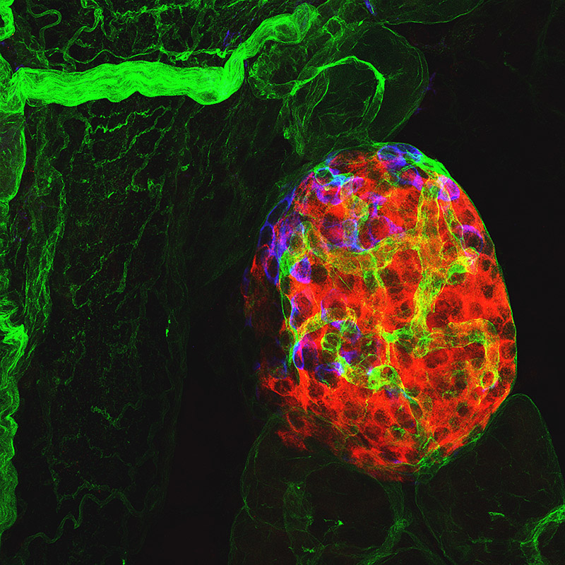
Cell clusters of the pancreas produce hormones that regulate blood sugar: alpha cells produce glucagon (blue); beta cells produce insulin (red). In type 1 diabetes, beta cells die and blood sugar becomes unbalanced. Confocal fluorescence microscopy© Eva Korpos, Lydia Sorokin 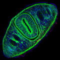
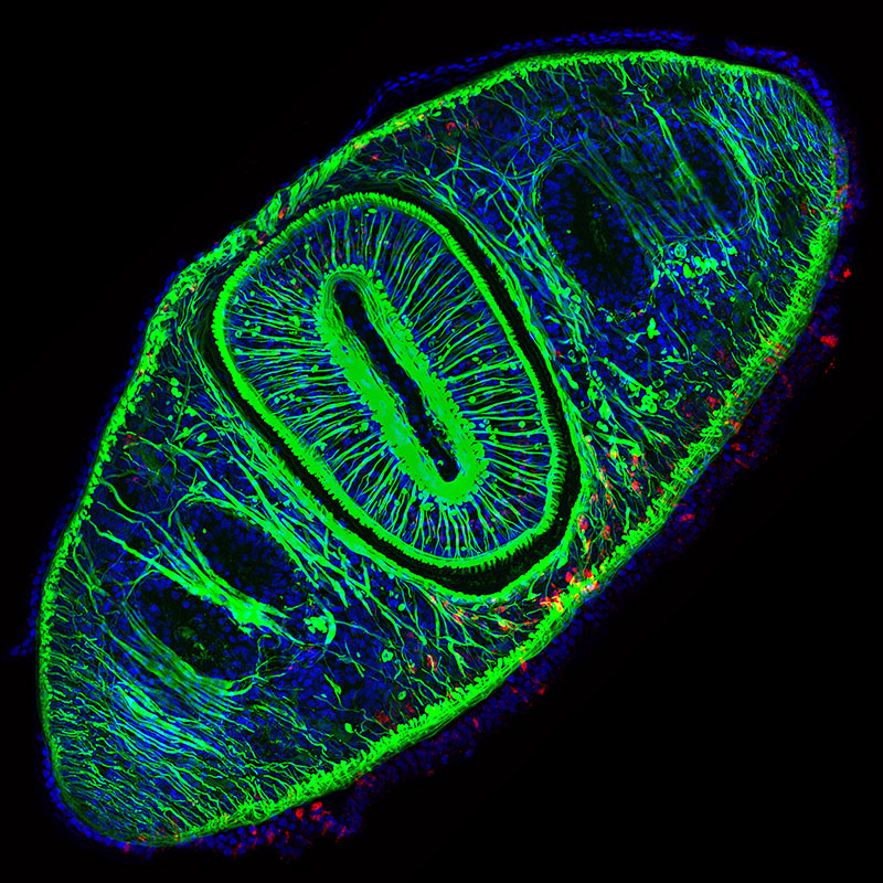
The flatworm is a master of regeneration. Thanks to a large number of stem cells, it can completely regenerate upon injury. Muscle cells (red) help in this process by sending signals to the stem cells. Confocal fluorescence microscopy© Florian Seebeck, Kerstin Bartscherer 
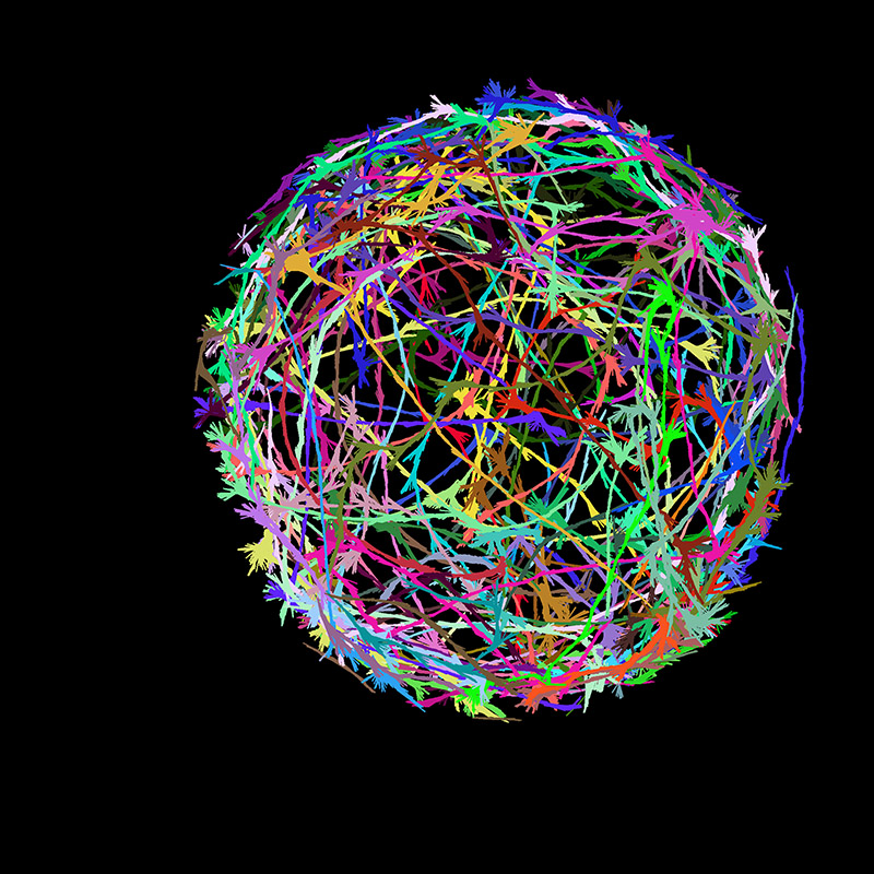
Paths of moving fruit fly larvae. Cellular processes in their brains influence their motion. Using a newly developed technique, CiM researchers study the behaviour of the tiny organisms. Each colour represents the motion of one larva. FIM imaging© Benjamin Risse, Nils Otto, Dimitri Berh, Xiaoyi Jiang, Christian Klämbt


A set of 27 colourful images from research in the Cells-in-Motion Cluster of Excellence give impressive insights into the inner workings of cells and organisms.